
Impact of gaze deviation on the optic nerve sheath diameter metric: A prospective interventional study
Keywords:
ONSD, Point of Care Ultrasound, Neurocritical care, Ocular ultrasonography, ONSD squeeze, 30-degree test, gazeAbstract
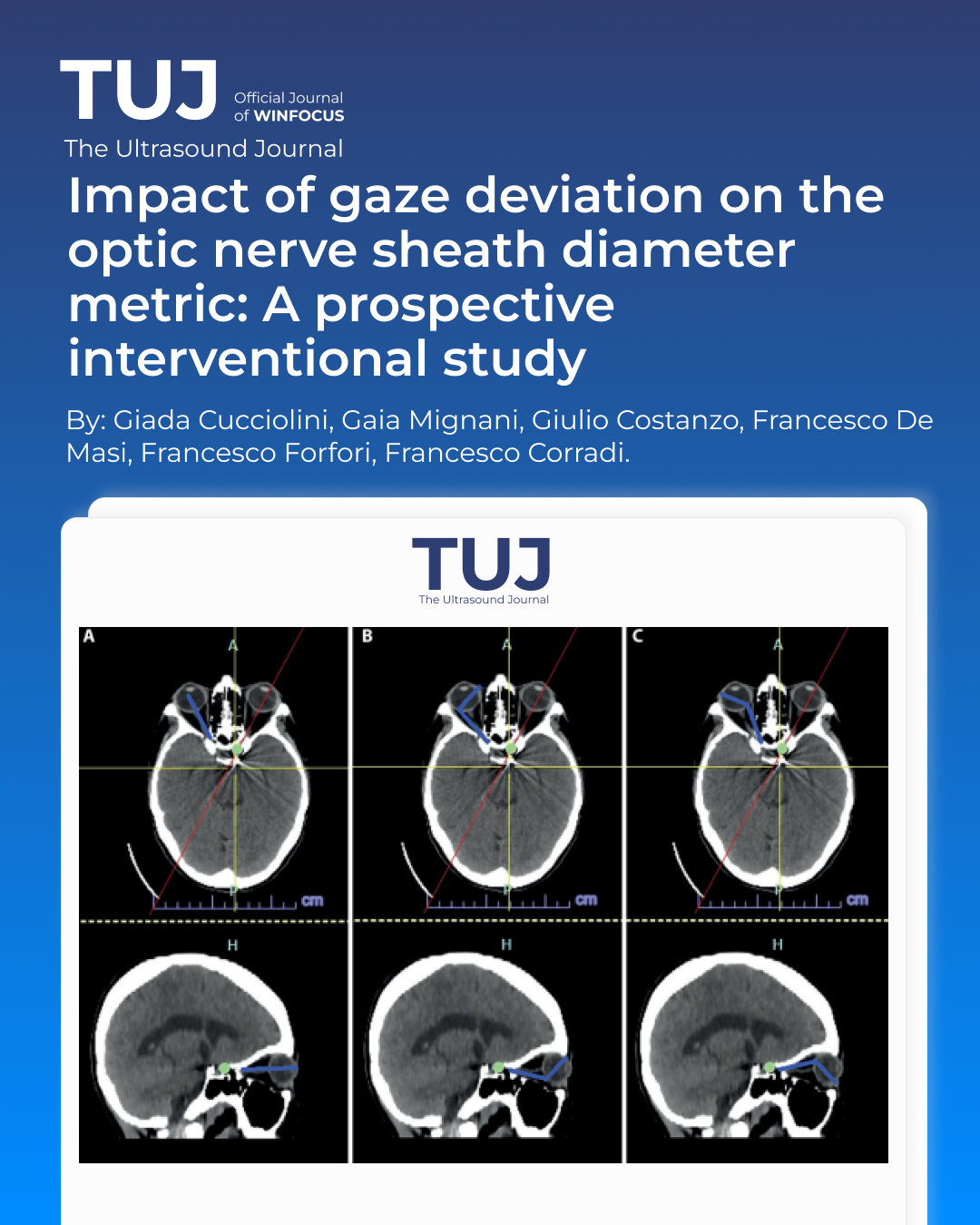
Background: Optic nerve sheath diameter (ONSD) is a widely used non-invasive surrogate marker of intracranial pressure. Current recommendations state that measurements should be obtained with a neutral gaze. However, the effect of gaze deviation on ONSD has never been investigated. We aimed to evaluate the reliability of ONSD in case of gaze deviation and quantify the associated measurement error. A secondary aim was to assess whether bed inclination has a significant effect on ONSD values.
Methods: We conducted a prospective interventional study on 44 healthy volunteers during routine ocular ultrasound training. ONSD was measured bilaterally along the horizontal and vertical axes at 45° head-of-bed elevation in neutral and deviated gaze. After repositioning the bed to 0° and a 2-minute stabilization period, horizontal measurements were repeated. Measurements were performed according to current consensus recommendations and ALARA principles. Nonparametric analyses were used to analyse the results.
Results: Gaze deviation significantly reduced ONSD values across all axes and both eyes, with a consistent median difference of 0.8–1.0 mm (all p < 0.001) and large effect sizes. In contrast, no significant differences were observed between measurements obtained at 45° and 0° (p > 0.5).
Conclusions: Gaze deviation induces a predictable “stretching” effect on the optic nerve sheath, resulting in clinically relevant ONSD reduction. If confirmed in neurocritical populations, this systematic difference may allow estimation of neutral-gaze ONSD in non compliant patients and could form the basis for a dynamic test that estimates optic nerve sheath compliance.
References
1. Hirzallah MI, Lochner P, Hafeez MU, Lee AG, Krogias C, Dongarwar D, et al. Optic Nerve Sheath Diameter Point-of-Care Ultrasonography Quality Criteria Checklist: An International Consensus Statement on Optic Nerve Sheath Diameter Imaging and Measurement*. Crit Care Med. 2024 Oct;52(10):1543. doi:10.1097/CCM.0000000000006345
2. Li R, Tang W, Li P, Huang Q, She J, Li S, et al. Automated measurement of optic nerve sheath diameter using ocular ultrasound video. Med Biol Eng Comput. 2025 Oct 14. doi:10.1007/s11517-025-03442-7 PubMed PMID: 41087787.
3. von Elm E, Altman DG, Egger M, Pocock SJ, Gøtzsche PC, Vandenbroucke JP, et al. The Strengthening the Reporting of Observational Studies in Epidemiology (STROBE) statement: guidelines for reporting observational studies. Lancet. 2007 Oct 20;370(9596):1453–7. doi:10.1016/S0140-6736(07)61602-X PubMed PMID: 18064739.
4. R Core Team. R: A Language and Environment for Statistical Computing [Internet]. Vienna, Austria: R Foundation for Statistical Computing; 2023. Available from: https://www.R-project.org/
5. Hirzallah MI, Sarwal A, Dentinger AM, Robba C, Valaikienė J, Lochner P, et al. Ultrasonographic Optic Nerve Sheath Diameter Technical Pitfalls and Imaging Artifacts. J Ultrasound Med Off J Am Inst Ultrasound Med. 2025 Feb 11. doi:10.1002/jum.16655 PubMed PMID: 39931745.
6. Meiburger KM, Naldi A, Michielli N, Coppo L, Fassbender K, Molinari F, et al. Automatic Optic Nerve Measurement: A New Tool to Standardize Optic Nerve Assessment in Ultrasound B-Mode Images. Ultrasound Med Biol. 2020 Jun;46(6):1533–44. doi:10.1016/j.ultrasmedbio.2020.01.034 PubMed PMID: 32147099.
7. Netteland DF, Aarhus M, Smistad E, Sandset EC, Padayachy L, Helseth E, et al. Noninvasive intracranial pressure assessment by optic nerve sheath diameter: Automated measurements as an alternative to clinician-performed measurements. Front Neurol. 2023;14:1064492. doi:10.3389/fneur.2023.1064492 PubMed PMID: 36816558; PubMed Central PMCID: PMC9928958.
8. Copetti R, Cattarossi L. Optic nerve ultrasound: artifacts and real images. Intensive Care Med. 2009 Aug 1;35(8):1488–9. doi:10.1007/s00134-009-1494-4
9. Rosa N, De Bernardo M, Di Stasi M, Cione F, Capaldo I. A-Scan Ultrasonographic Evaluation of Patients with Idiopathic Intracranial Hypertension: Comparison of Optic Nerves. J Clin Med. 2022 Oct 19;11(20):6153. doi:10.3390/jcm11206153 PubMed PMID: 36294473; PubMed Central PMCID: PMC9605245.
10. Pardon LP, Cheng H, Chettry P, Patel NB. Optic Nerve Head Morphological Changes Over 12 Hours in Seated and Head-Down Tilt Postures. Invest Ophthalmol Vis Sci. 2020 Nov 2;61(13):21. doi:10.1167/iovs.61.13.21 PubMed PMID: 33186468; PubMed Central PMCID: PMC7671873.
11. Ustick JJ, Pardon LP, Chettry P, Patel NB, Cheng H. Effects of Head-Down Tilt on Optic Nerve Sheath Diameter in Healthy Subjects. Ophthalmic Physiol Opt J Br Coll Ophthalmic Opt Optom. 2023 Nov;43(6):1531–9. doi:10.1111/opo.13200 PubMed PMID: 37401194; PubMed Central PMCID: PMC10592427.
Downloads
Published
Issue
Section
License
Copyright (c) 2026 Giada Cucciolini, Gaia Mignani, Giulio Costanzo, Francesco De Masi, Francesco Forfori, Francesco Corradi (Author)

This work is licensed under a Creative Commons Attribution-NonCommercial 4.0 International License.
This is an Open Access article distributed under the terms of the Creative Commons Attribution License (https://creativecommons.org/licenses/by-nc/4.0) which permits unrestricted use, distribution, and reproduction in any medium, provided the original work is properly cited.
Authors retain the copyright for their published work. No formal permission will be required to reproduce parts (tables or illustrations) of published papers, provided the source is quoted appropriately and reproduction has no commercial intent.
How to Cite


