
Estimate the severity of acute ischemic stroke by optic nerve sheath ultrasound
Keywords:
Ultrasound, Optic nerve sheath diameter, Stroke, Severity, ICPAbstract
Background: Timely diagnosis of acute ischemic stroke can aid optimal treatment. Optic nerve sheath diameter (ONSD) can determine increased intracranial pressure (ICP) in such cases. The purpose of this study is to determine the value of ONSD in estimating the severity of acute ischemic stroke.
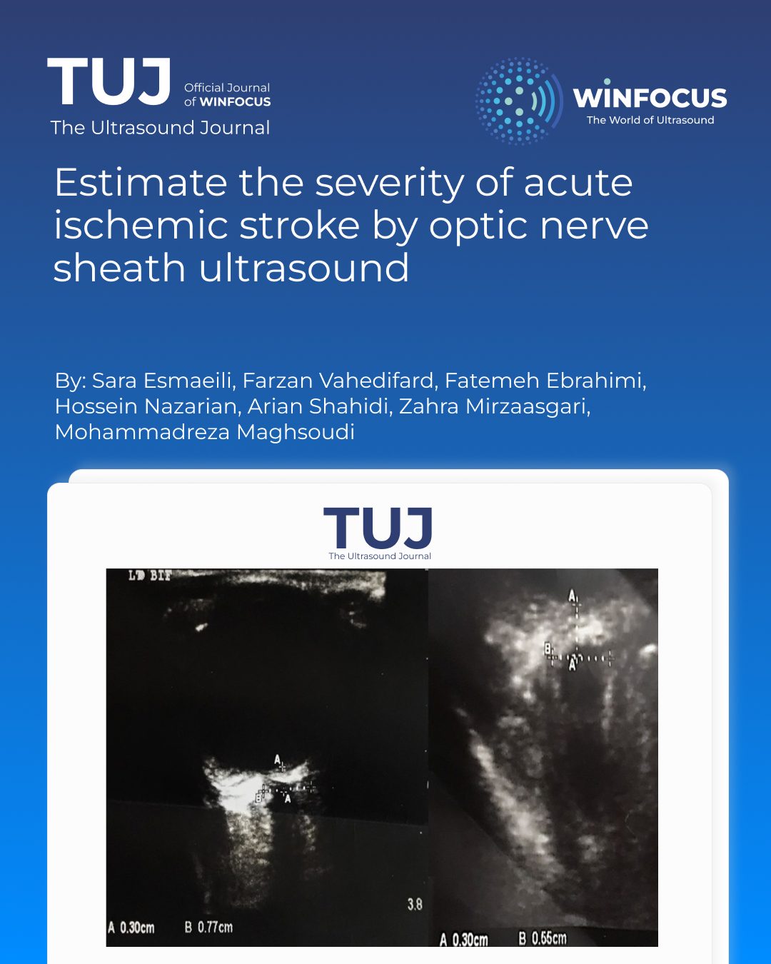
Methods: Patients with acute ischemic stroke who were referred to a stroke center were studied. The ONSD of both the right and left sides was measured by ultrasound on the day of admission. Ischemic stroke severity was determined based on the NIHSS.
Results: A strong correlation was found between increased right and left ONSDs and severity of ischemic stroke determined by the initial NIHSS score. Based on ROC curve (receiver operating characteristic curve) analysis, both cut points of 5.65 mm for right ONSD (with 100% sensitivity of and 86% specificity) and 5.75 mm for left ONSD (with a 100% Sensitivity and 88% specificity) were able to predict severe stroke. The value of the right ONSD (Area Under the Curve = 0.959) and the left ONSD (Area Under the Curve = 0.942) indicated a strong predictive value.
Conclusions: Ultrasound as a feasible and non-invasive modality might play a role in determining the severity of an acute ischemic stroke, and could be considered a promising first-line decision making tool.
References
1. Deljavan R, Farhoudi M, Sadeghi-Bazargani H (2018) Stroke in-hospital survival and its predictors: the first results from Tabriz stroke registry of Iran. Int J Gen Med 11:233–240
2. Michinaga S, Koyama Y (2015) Pathogenesis of brain edema and investigation into anti-edema drugs. Int J Mol Sci 16(5):9949–9975
3. Dostovic Z, Dostovic E, Smajlovic D, Ibrahimagic OC, Avdic L (2016) Brain edema after ischaemic stroke. Med Arch (Sarajevo, Bosnia and Herzegovina) 70(5):339 –41
4. Campbell BCV, De Silva DA, Macleod MR, Coutts SB, Schwamm LH, Davis SM et al (2019) Ischaemic stroke. Nat Reviews Disease Primers 5(1):70
5. Regenhardt RW, Das AS, Stapleton CJ, Chandra RV, Rabinov JD, Patel AB et al (2017) Blood pressure and penumbral sustenance in stroke from large vessel occlusion. Front Neurol 8:317
6. Kinoshita K (2016) Traumatic brain injury: pathophysiology for neurocritical care. J Intensive Care 4:29
7. Roh D, Park S (2016) Brain multimodality monitoring: updated perspectives. Curr Neurol Neurosci Rep 16(6):56
8. Treggiari MM, Schutz N, Yanez ND, Romand JA (2007) Role of intracranial pressure values and patterns in predicting outcome in traumatic brain injury: a systematic review. Neurocrit Care 6(2):104–112
9. Czosnyka M, Pickard JD, Steiner LA (2017) Principles of intracranial pressure monitoring and treatment. Handb Clin Neurol 140:67–89
10. Tripathy S, Ahmad SR (2019) Raised intracranial pressure syndrome: A Stepwise Approach. Indian journal of critical care medicine: peer-reviewed. Official Publication Indian Soc Crit Care Med 23(Suppl 2):S129–s35
11. Malayeri AA, Bavarian S, Mehdizadeh M (2005) Sonographic evaluation of optic nerve diameter in children with Raised intracranial pressure. J Ultrasound Medicine: Official J Am Inst Ultrasound Med 24(2):143–147
12. Harary M, Dolmans RGF, Gormley WB (2018) Intracranial pressure monitoring-review and avenues for development. Sensors 18(2)
13. Khan MN, Shallwani H, Khan MU, Shamim MS (2017) Noninvasive monitoring intracranial pressure - A review of available modalities. Surg Neurol Int 8:51
14. Hanafi MG, Verki MM, Parei SN (2019) Ultrasonic assessment of optic nerve sheath to detect increased intracranial pressure. J Med Ultrasound 27(2):69–74
15. Wang J, Li K, Li H, Ji C, Wu Z, Chen H et al (2019) Ultrasonographic optic nerve sheath diameter correlation with ICP and accuracy as a tool for noninvasive surrogate ICP measurement in patients with decompressive craniotomy. J Neurosurg 1–7
16. Adams HP, Bendixen BH, Kappelle LJ, Biller J, Love BB, Gordon DL et al (1993) Classification of subtype of acute ischemic stroke. Definitions for use in a multicenter clinical trial. TOAST. Trial of org 10172 in acute stroke treatment. Stroke 24(1):35–41
17. Gökcen E, Caltekin İ, Savrun A, Korkmaz H, Savrun ŞT, Yıldırım G (2017) Alterations in optic nerve sheath diameter according to cerebrovascular disease sub-groups. Am J Emerg Med 35(11):1607–1611
18. Hedna VS, Rastogi V, Weeks E, Patel RJS (2015) Abstract W MP83: use of optic nerve sheath diameter in emergency department to predict. Stroke Outcome 46
19. Komut E, Kozacı N, Sönmez BM, Yılmaz F, Komut S, Yıldırım ZN et al (2016) Bedside sonographic measurement of optic nerve sheath diameter as a predictor of intracranial pressure in ED. Am J Emerg Med 34(6):963–967
20. Aduayi OS, Asaleye CM, Adetiloye VA, Komolafe EO, Aduayi VA (2015) Optic nerve sonography: A noninvasive means of detecting Raised intracranial pressure in a resource-limited setting. J Neurosciences Rural Pract 6(4):563–567
21. Jeon JP, Lee SU, Kim SE, Kang SH, Yang JS, Choi HJ, et al. Correlation of optic nerve sheath diameter with directly measured intracranial pressure in Korean adults using bedside ultrasonography. PloS one. 2017;12(9):e0183170.
22. Wang L, Feng L, Yao Y, Wang Y, Chen Y, Feng J, et al. Optimal Optic Nerve Sheath Diameter Threshold for the Identification of Elevated Opening Pressure on Lumbar Puncture in a Chinese Population. PloS one. 2015;10(2):e0117939
23. Munawar K, Khan MT, Hussain SW, Qadeer A, Shad ZS, Bano S, et al. Optic Nerve Sheath Diameter Correlation with Elevated Intracranial Pressure Determined via Ultrasound. Cureus. 2019;11(2):e4145.
24. Liu KC, Fleischman D, Lee AG, Killer HE, Chen JJ, Bhatti MT. Current concepts of cerebrospinal fluid dynamics and the translaminar cribrosa pressure gradient: a paradigm of optic disk disease. Survey of Ophthalmology. 2020;65(1):48-66.
25. Moodley AA, Dlwati MS, Durand M. Intracanalicular Optic Nerve Swelling and Signal Change in Fulminant Untreated Idiopathic Intracranial Hypertension. Neuro-ophthalmology (Aeolus Press). 2017;41(2):84-9.
Downloads
Published
Issue
Section
License
Copyright (c) 2025 Sara Esmaeili, Farzan Vahedifard, Fatemeh Ebrahimi, Hossein Nazarian, Arian Shahidi, Zahra Mirzaasgari, Mohammadreza Maghsoudi (Author)

This work is licensed under a Creative Commons Attribution-NonCommercial 4.0 International License.
This is an Open Access article distributed under the terms of the Creative Commons Attribution License (https://creativecommons.org/licenses/by-nc/4.0) which permits unrestricted use, distribution, and reproduction in any medium, provided the original work is properly cited.
Authors retain the copyright for their published work. No formal permission will be required to reproduce parts (tables or illustrations) of published papers, provided the source is quoted appropriately and reproduction has no commercial intent.
How to Cite


