
Performance evaluation of the TE Air wireless handheld ultrasound in cardiac applications: a prospective comparative study
Keywords:
Handheld ultrasound, Wireless, TE Air, Cardiac ultrasoundAbstract
Aim: To evaluate the reliability and reproducibility of the TE Air wireless handheld ultrasound device in clinical cardiac imaging by comparing its performance with a high-end reference system.
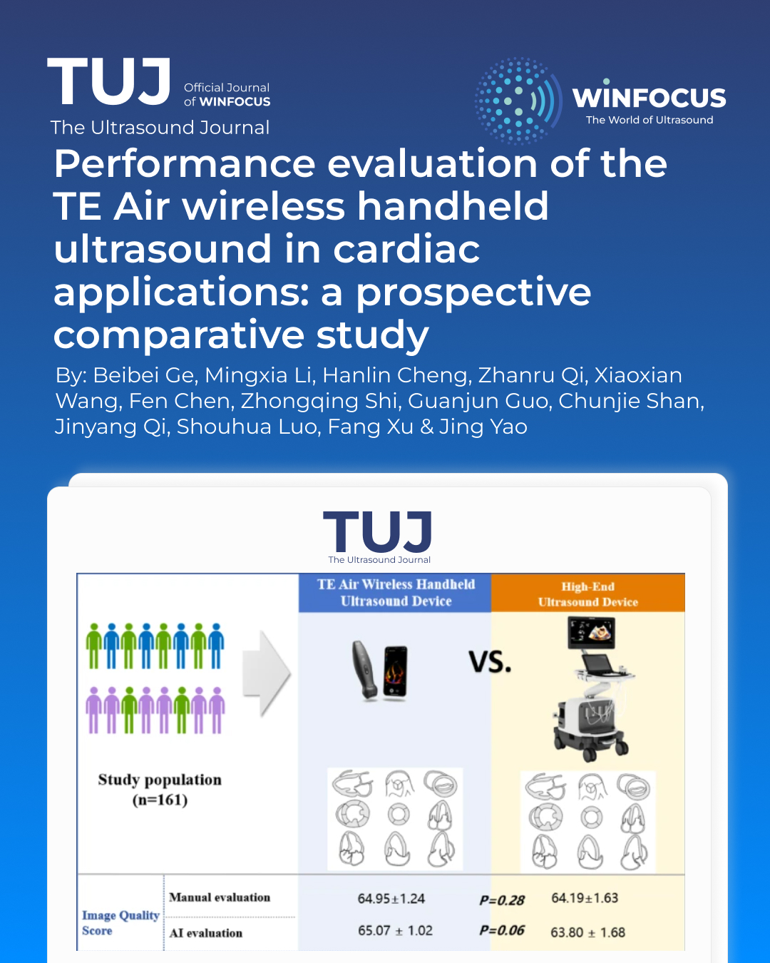
Methods: 161 patients for good-quality echocardiographic images were included in this prospective study. Each patient underwent sequential imaging using both the TE Air device (Mindray) and the high-end reference device (Philips EPIQ 7 C). Nine standard cardiac views were acquired. Image quality was assessed manually by two blinded echocardiographers and via proprietary AI software, respectively. The following key parameters were analyzed basing on the images: diastolic thickness of interventricular septal (IVSTd) and left ventricular posterior wall (LVPWTd), left ventricular end-diastolic (LVDd) and end-systolic diameter (LVDs), aortic diameter (AOD), left atrial anteroposterior diameter (LAD), Early (E) and late (A) diastolic velocities of the mitral valve in PW mode, as well as early diastolic velocities at the septal (EmS) and lateral (EmL) mitral annulus. Regional wall motion abnormality (RWMA), bicuspid aortic valve (BAV), atrial septal defect (ASD), left ventricular ejection fraction (LVEF) and valvular regurgitation degree were independently evaluated.
Results: The TE Air demonstrated comparable image quality to the high-end reference system in both manual (64.95 ± 1.24 vs. 64.19 ± 1.63, P = 0.28) and AI-based evaluations (65.07 ± 1.02 vs. 63.80 ± 1.68, P = 0.06). Structural measurements showed high inter-device consistency, with ICCs of 0.77/0.74 for IVSTd/LVPWTd, 0.95/0.96 for LVDd/LVDs, and 0.82/0.98 for AOD/LAD (all P < 0.001). Functional parameters also demonstrated strong agreement (ICC: 0.91/0.92 for mitral E/A waves; 0.79/0.85 for EmS/EmL; P < 0.001). The TE Air had sensitivities of 81.8% for RWMA, 100% for ASD and BAV, and 93.5% for LVEF < 50%. Diagnostic agreement was excellent for LVEF (κ = 0.96, P < 0.001) and valvular regurgitation (weighted κ = 0.89, P < 0.001).
Conclusion: The TE Air wireless handheld ultrasound device exhibits high agreement with high-end reference device in image quality, measurements, and clinical diagnoses, supporting its potential for widespread use in point-of-care ultrasound (POCUS) clinical applications.
References
1. Ben-Baruch Golan Y, Sadeh R, Mizrakli Y et al (2020) Early Point-of-Care ultrasound assessment for medical patients reduces time to appropriate treatment: A pilot randomized controlled trial. Ultrasound Med Biol 46:1908–1915. https://doi.org/10.1016/j.ultrasmedbio.2020.03.023
2. Díaz-Gómez JL, Mayo PH, Koenig SJ (2021) Point-of-Care ultrasonography. N Engl J Med 385:1593–1602. https://doi.org/10.1056/NEJMra1916062
3. Kalagara H, Coker B, Gerstein NS et al (2022) Point-of-Care ultrasound (POCUS) for the cardiothoracic anesthesiologist. J Cardiothorac Vasc Anesth 36:1132–1147. https://doi.org/10.1053/j.jvca.2021.01.018
4. Yoshida T, Yoshida T, Noma H, Nomura T, Suzuki A, Mihara T (2023) Diagnostic accuracy of point-of-care ultrasound for shock: a systematic review and meta-analysis. Crit Care 27:200. https://doi.org/10.1186/s13054-023-04495-6
5. Stock KF, Klein B, Steubl D et al (2015) Comparison of a pocket-size ultrasound device with a premium ultrasound machine: diagnostic value and time required in bedside ultrasound examination. Abdom Imaging 40:2861–2866. https://doi.org/10.1007/s00261-015-0406-z
6. Thavanathan Rajiv S, Woo Michael Y, Greg H (2020) The future is in your hands - Handheld ultrasound in the emergency department. World Rev Nutr Diet 22:55–59. https://doi.org/10.1159/000516732
7. Acuña J, Situ-LaCasse E, Yarnish AA, McNinch NL, Adhikari S (2024) Does size matter? A prospective study on the feasibility of using a handheld ultrasound device in place of a Cart-Based system in the evaluation of trauma patients. J Emerg Med 66:e483–e491. https://doi.org/10.1016/j.jemermed.2023.11.012
8. Jenkins S, Alabed S, Swift A et al (2021) Diagnostic accuracy of handheld cardiac ultrasound device for assessment of left ventricular structure and function: systematic review and meta-analysis. Heart 107:1826–1834. https://doi.org/10.1136/heartjnl-2021-319561
9. Rykkje A, Carlsen JF, Nielsen MB (2019) Hand-Held ultrasound devices compared with High-End ultrasound systems: A systematic review. Diagnostics (Basel) 9:61. https://doi.org/10.3390/diagnostics9020061
10. Busch M (2006) Portable ultrasound in pre-hospital emergencies: a feasibility study. Acta Anaesthesiol Scand 50:754–758. https://doi.org/10.1111/j.1399-6576.2006.01030.x
11. Lapostolle F, Petrovic T, Lenoir G et al (2006) Usefulness of hand-held ultrasound devices in out-of-hospital diagnosis performed by emergency physicians. Am J Emerg Med 24:237–242. https://doi.org/10.1016/j.ajem.2005.07.010
12. Jung EM, Dinkel J, Verloh N et al (2021) Wireless point-of-care ultrasound: first experiences with a new generation handheld device. Clin Hemorheol Microcirc 79:463–474. https://doi.org/10.3233/CH-211197
13. Newhouse SM, Effing TW, Dougherty BD, D’Costa JA, Rose AR (2020) Is bigger really better? Comparison of ultraportable handheld ultrasound with standard Point-of-Care ultrasound for evaluating safe site identification and image quality prior to pleurocentesis. Respiration 99:325–332. https://doi.org/10.1159/000505698
14. Zardi EM, Franceschetti E, Giorgi C, Palumbo A, Franceschi F (2019) Accuracy and performance of a new handheld ultrasound machine with wireless system. Sci Rep 9:14599. https://doi.org/10.1038/s41598-019-51160-6
15. Jung EM, Jung F, Dong Y, Kaiser U (2024) Initial description of the novel handheld wireless ultrasound device TE air with doppler and color duplex imaging. Clin Hemorheol Microcirc 86:89–97. https://doi.org/10.3233/CH-238100
16. Perez-Sanchez A, Johnson G, Pucks N et al (2024) Comparison of 6 handheld ultrasound devices by point-of-care ultrasound experts: a cross-sectional study. Ultrasound J 16:45. https://doi.org/10.1186/s13089-024-00392-3
17. Mitchell C, Rahko PS, Blauwet LA et al (2019) Guidelines for performing a comprehensive transthoracic echocardiographic examination in adults: recommendations from the American society of echocardiography. J Am Soc Echocardiogr 32:1–64. https://doi.org/10.1016/j.echo.2018.06.004
18. McDonagh TA, Metra M, Adamo M et al (2021) 2021 ESC guidelines for the diagnosis and treatment of acute and chronic heart failure. Eur Heart J 42:3599–3726. https://doi.org/10.1093/eurheartj/ehab368
19. Lancellotti P, Pibarot P, Chambers J et al (2022) Multi-modality imaging assessment of native valvular regurgitation: an EACVI and ESC Council of valvular heart disease position paper. Eur Heart J Cardiovasc Imaging 23:e171–e232. https://doi.org/10.1093/ehjci/jeab253
20. Lancellotti P, Tribouilloy C, Hagendorff A et al (2013) Recommendations for the echocardiographic assessment of native valvular regurgitation: an executive summary from the European association of cardiovascular imaging. Eur Heart J Cardiovasc Imaging 14:611–644. https://doi.org/10.1093/ehjci/jet105
Downloads
Published
Issue
Section
License
Copyright (c) 2025 Beibei Ge, Mingxia Li, Hanlin Cheng, Zhanru Qi, Xiaoxian Wang, Fen Chen, Zhongqing Shi, Guanjun Guo, Chunjie Shan, Jinyang Qi, Shouhua Luo, Fang Xu, Jing Yao (Author)

This work is licensed under a Creative Commons Attribution-NonCommercial 4.0 International License.
This is an Open Access article distributed under the terms of the Creative Commons Attribution License (https://creativecommons.org/licenses/by-nc/4.0) which permits unrestricted use, distribution, and reproduction in any medium, provided the original work is properly cited.
Authors retain the copyright for their published work. No formal permission will be required to reproduce parts (tables or illustrations) of published papers, provided the source is quoted appropriately and reproduction has no commercial intent.
How to Cite


