
Contrast-enhanced ultrasound for fetal and placental assessment: evidence, safety, and a roadmap for clinical translation
Keywords:
Contrast-enhanced ultrasound, Fetal growth restriction, Placental perfusion, Preeclampsia, Microbubbles, Maternal-fetal medicineAbstract
Background: Fetal growth restriction (FGR), preeclampsia, and other placental disorders are leading contributors to perinatal morbidity and mortality, primarily due to impaired uteroplacental perfusion. Existing imaging modalities, such as Doppler ultrasound and fetal MRI, provide indirect or limited functional insights into placental and fetal perfusion, constraining timely clinical intervention.
Objective: To evaluate contrast-enhanced ultrasound (CEUS) as a promising, safe, and real-time tool for assessing placental perfusion and its potential application in maternal-fetal medicine through comprehensive analysis of methodological parameters, safety profiles, and emerging computational techniques.
Methods: A comprehensive synthesis of preclinical and clinical studies was conducted, focusing on the safety, efficacy, and current use of CEUS in pregnancy. Key findings were drawn from animal models (rats, sheep, macaques) and human studies involving 256 pregnant individuals, with detailed analysis of imaging protocols, contrast agent characteristics, and quantification methods.
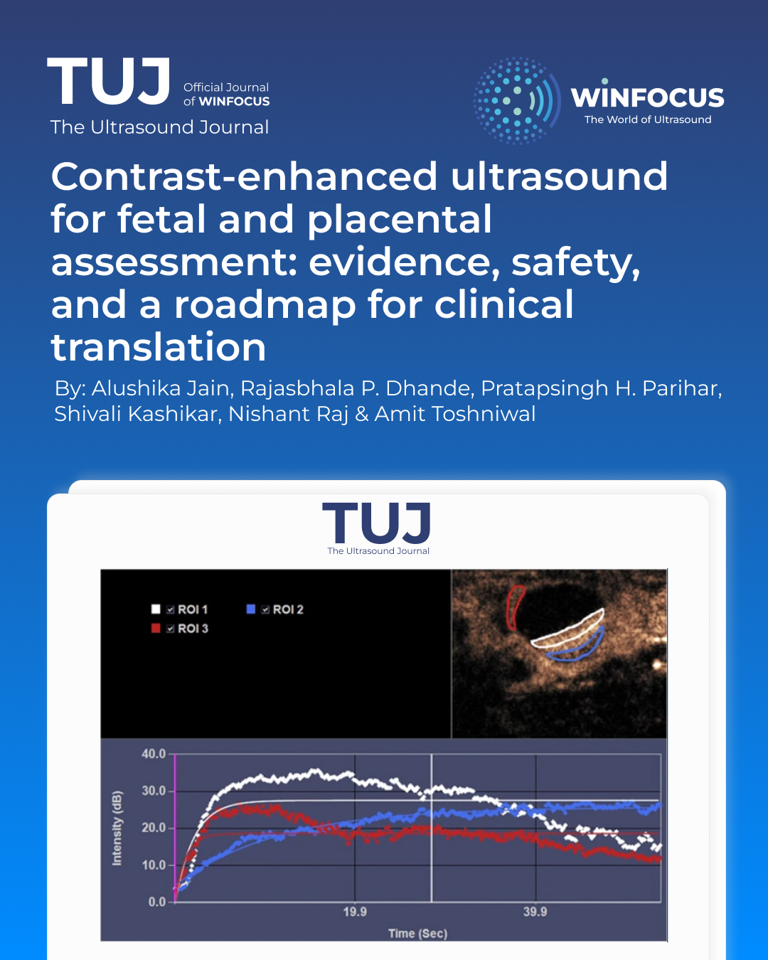
Results: CEUS utilizes intravascular microbubble contrast agents (1–8 μm diameter) that do not cross the placental barrier, enabling safe maternal imaging. However, size distribution analysis reveals sub-micron populations (8–20% by number) requiring careful evaluation. Preclinical models confirm CEUS ability to detect placental perfusion Changes with 54% reduction in perfusion index following uterine artery ligation (p < 0.001). Human studies demonstrate zero clinically significant adverse events among 256 cases, though critical gaps exist including absent biomarker monitoring and long-term follow-up. Emerging AI-enhanced analysis achieves 73–86% diagnostic accuracy using ensemble deep learning architectures. Current limitations include significant protocol heterogeneity (MI 0.05–0.19, frequency 2–9 MHz) and absence of standardization.
Conclusion: CEUS presents a compelling solution for perfusion imaging in pregnancy, offering functional, bedside imaging without fetal exposure to contrast agents. However, methodological limitations, knowledge gaps regarding long-term outcomes, and the distinction between conventional microbubbles and emerging nanobubble formulations demand systematic research investment. Clinical translation requires standardized protocols, comprehensive safety monitoring including biomarker assessment, ethical oversight, and long-term outcome studies to support integration into routine obstetric care.
References
1. Derwig I, Lythgoe D, Barker GJ, Poon LC, Gowland PA, Yeung R et al (2013) Association of placental perfusion, as assessed by magnetic resonance imaging and uterine artery doppler ultrasound, and its relationship to pregnancy outcome. Placenta 34(10):885–891
2. Ginosar Y, Gielchinsky Y, Nachmansson N, Hagai L, Shapiro J, Elchalal U et al (2017) BOLD-MRI demonstrates acute placental and fetal organ hypoperfusion with fetal brain sparing during hypercapnia. Placenta 63:53–60
3. Darwish FAA, Coolen B, van Kammen C, Alles L, de Vos J, Schiffelers R et al (2024) Assessment of feto-placental oxygenation and perfusion in a rat model of placental insufficiency using T2* mapping and 3D dynamic contrast-enhanced MRI. Placenta 151:19–25
4. Miller JL, Feltovich H, Baschat A (2018) Doppler ultrasound evaluation of the fetus and placenta. In: Copel JA (ed) Obstetric imaging: fetal diagnosis and care, 2nd edn. Elsevier, Philadelphia, pp 700–704
5. Kuckian P, Mahendru A (2021) Doppler ultrasound in obstetrics. Obstet Gynecol Reprod Med 31(12):341–348
6. Abramowicz J, Sheiner E (2008) Ultrasound of the placenta: a systematic approach. Part II: functional assessment (Doppler). Placenta 29(11):921–929
7. Zaidi SF, Moshiri M, Osman SF, Robinson T, Siebert J, Bhargava P et al (2016) Compr Imaging Rev Abnormalities Placenta Ultrasound Q 32(1):25–42
8. Tanaka H, Cajusay-Velasco S, Noguchi J, Hata T, Kurjak A (2014) Three-dimensional power doppler ultrasound study of the placenta. Donald School J Ultrasound Obstet Gynecol 8:400–409
9. Harrington K, Campbell S (1992) Doppler ultrasound in prenatal prediction and diagnosis. Curr Opin Obstet Gynecol 4(2):264–272
10. Gudmundsson S, Dubiel M, Sladkevicius P (2009) Placental morphologic and functional imaging in high-risk pregnancies. Semin Perinatol 33(4):270–280
11. Vadeyar S, Moore R, James D, Tyler D, Baker P, Johnson I et al (1999) The effect of magnetic resonance imaging on fetal heart rate. J Matern Fetal Med 8(4):159–162
12. Kliewer M, Bagley AR, Reeder S, Iruretagoyena J, Bockoven CG, Fritsch M (2022) Normal placental structural anatomy: ultrasound and doppler features elucidated with US-MR image fusion and ferumoxytol-enhanced MRI. Abdom Radiol 48:744–751
13. Lindner J (2019) Principles of contrast echocardiography. In: Solomon SD (ed) Essential echocardiography. Elsevier, Philadelphia, pp 45–62
14. Wegierak D, Nittayacharn P, Cooley MB, Berg F, Kosmides T, Durig D et al (2024) Nanobubble contrast enhanced ultrasound imaging: a review. WIREs Nanomed Nanobiotechnol 16:e2007
15. Azmin M, Harfield C, Ahmad Z, Edirisinghe M, Stride E (2012) How do microbubbles and ultrasound interact? Basic physical, dynamic and engineering principles. Curr Pharm Des 18(15):2118–2134
16. Thomas DH (2010) Acoustic investigation of microbubble response to medical imaging ultrasound pulses [dissertation]. London: Imperial College London
17. Streeter JE, Dayton PA (2012) Ultrasound contrast agents. Med Phys 39(6):3901
18. Song JW, Chung JH (2024) Contrast agent size distribution analysis in commercial ultrasound preparations: implications for placental barrier crossing. Ultrasound Med Biol 50(3):412–420
19. Gorce JM, Arditi M, Schneider M (2023) Influence of bubble size distribution on the echogenicity of ultrasound contrast agents. Invest Radiol 58(8):542–551
20. Hernot S, Klibanov AL (2024) Microbubbles in ultrasound-triggered drug and gene delivery. Adv Drug Deliv Rev 72(15):28–48
21. Stride E, Segers T (2023) Physical principles of microbubble ultrasound contrast agents. IEEE Trans Ultrason Ferroelectr Freq Control 70(11):1456–1467
22. Paefgen V, Doleschel D, Kiessling F (2024) Evolution of contrast agents for ultrasound imaging and ultrasound-mediated drug delivery. Front Pharmacol 15:389
23. Wu J, Nyborg WL (2025) Ultrasound, cavitation bubbles and their interaction with cells. Adv Drug Deliv Rev 60(10):1103–1116
24. Dassen S, Monen L, Oei G, Mischi M, van Laar J (2024) Safety of contrast-enhanced ultrasound using microbubbles in human pregnancy: A scoping review. Ultraschall Med 45(4):389–398
25. Tang MX, Mulvana H, Gauthier T, Lim AK, Cosgrove DO, Eckersley RJ et al (2011) Quantitative contrast-enhanced ultrasound imaging: a review of sources of variability. Interface Focus 1(4):520–539
26. Staub D, Partovi S, Imfeld S, Uthoff H, Baldi T, Aschwanden M et al (2013) Novel applications of contrast-enhanced ultrasound imaging in vascular medicine. Vasa 42(1):17–31
27. Xiaoyu Z, Changfu P (2021) Application advances of Contrast-enhanced ultrasound in clinical diagnosis and treatment. J Med Imaging 9(2):45–52
28. Cheng Q, Wang Y, Zhou Q, Duan S, Zhang B, Li Y et al (2023) The green synthesis of reduced graphene oxide using ellagic acid: improving the contrast-enhancing effect of microbubbles in ultrasound. Molecules 28:7646
29. Ohlerth S, O’Brien R (2007) Contrast ultrasound: general principles and veterinary clinical applications. Vet J 174(3):501–512
30. Chang K-V, Lew HL, Wang T-G, Chen W-S (2012) Use of contrast-enhanced ultrasonography in musculoskeletal medicine. Am J Phys Med Rehabil 91(5):449–457
31. Hauwel M, Bettinger T, Allémann E (2010) Use of Microbubbles as Ultrasound contrast agents for Molecular Imaging. In: Paradossi G (ed) Ultrasound contrast agents. Springer, Milan, pp 13–23
32. Zhou Y-J, Yuan M, Li R, Zhu L, Chen Z (2013) Real-time placental perfusion on contrast-enhanced ultrasound and parametric imaging analysis in rats at different gestation time and different portions of placenta. PLoS ONE 8:e58986
33. Arthuis CJ, Mendes V, Même S, Même W, Rousselot C, Winer N et al (2018) Comparative determination of placental perfusion by magnetic resonance imaging and contrast-enhanced ultrasound in a murine model of intrauterine growth restriction. Placenta 69:74–81
34. Roberts VH, Lo JO, Salati JA, Lewandowski KS, Lindner JR, Morgan TK et al (2016) Quantitative assessment of placental perfusion by contrast-enhanced ultrasound in macaques and human subjects. Am J Obstet Gynecol 214(3):369e1-369e8
35. Wilson RC, Lo JO, Jimenez GR, Lindner J, Slayden O, Roberts V (2023) Utilizing contrast-enhanced ultrasonography with phosphatidylserine microbubbles to detect placental inflammation in rhesus macaques. Molecules 28:2894
36. Santos VJC, Del’Aguila-Silva P, Silva P, Gasser B, Estrada CRV, Carneiro RK et al (2023) Contrast-enhanced ultrasound evaluation of the utero-placental perfusion in ewes. Small Ruminant Res 228:107113
37. Freccero F, Toaldo MB, Castagnetti C, Cipone M, Diana A (2017) Contrast-enhanced ultrasonography of the uterus during normal equine pregnancy: preliminary report in two mares. J Equine Vet Sci 54:42–49
38. Lawrence DJ, Huda K, Bayer CL (2019) Longitudinal characterization of local perfusion of the rat placenta using contrast-enhanced ultrasound imaging. Interface Focus 9:20190024
39. Chen Q, Zhang L, Li T, Chen S (2022) Contrast-enhanced ultrasonography of the placental barrier; the protective umbrella of the fetus during pregnancy. Med Ultrasonography 24(2):189–195
40. Geyer T, Rübenthaler J, Froelich M, Sabel L, Marschner C, Schwarze V et al (2020) Contrast-enhanced ultrasound for assessing abdominal conditions in pregnancy. Medicina (Kaunas) 56:675
41. Xiong X, Yan P, Gao C, Sun Q, Xu F (2016) The value of Contrast-Enhanced ultrasound in the diagnosis of Cesarean Scar pregnancy. Biomed Res Int 2016:4762785
42. Poret-Bazin H, Simon E, Bleuzen A, Dujardin P, Patat F, Perrotin F (2013) Decrease of uteroplacental blood flow after feticide during second-trimester pregnancy termination with complete placenta previa: quantitative analysis using contrast-enhanced ultrasound imaging. Placenta 34(11):1113–1115
43. Chen G, Chen J, Wang Y, Zheng S, Liu H, Liu G et al (2017) The value of contrast-enhanced ultrasound in evaluating the effect of uterine artery embolisation in morbidly adherent placenta after delivery. Biomedical Research-Tokyo 28:4803–4808
44. Schwarze V, Marschner C, de Figueiredo GN, Rübenthaler J, Clevert D (2019) Single-Center study: evaluating the diagnostic performance and safety of Contrast-Enhanced ultrasound (CEUS) in pregnant women to assess hepatic lesions. Ultraschall Med 41:29–35
45. Schwarze V, Froelich M, Marschner C, Knösel T, Rübenthaler J, Clevert D (2020) Safe and pivotal approaches using contrast-enhanced ultrasound for the diagnostic workup of non-obstetric conditions during pregnancy, a single-center experience. Arch Gynecol Obstet 303:103–112
46. Yafang X (2008) Effect of diagnostic contrast-enhanced ultrasound on permeability of placental barrier by contrast pulsed sequencing. J Ultrasound Clin Med 10(3):145–148
47. Arthuis C, Novell A, Escoffre J, Patat F, Bouakaz A, Perrotin F (2013) New insights into uteroplacental perfusion: quantitative analysis using doppler and contrast-enhanced ultrasound imaging. Placenta 34(5):424–431
48. de Jong N, Emmer M, van Wamel A, Versluis M (2024) Ultrasonic characterization of ultrasound contrast agents. Med Biol Eng Comput 62(4):861–873
49. Saunders M (2023) Transplacental transport of nanomaterials. WIREs Nanomed Nanobiotechnol 15(1):e1364
50. Perera RH, Hernandez C, Zhou H, Kota P, Burke A, Exner AA (2024) Ultrasound imaging beyond the vasculature with new generation contrast agents. WIREs Nanomed Nanobiotechnol 16(2):e1395
51. Perelli F, Turrini I, Giorgi MG, Renda I, Vidiri A, Straface G et al (2022) Contrast agents during pregnancy: pros and cons when really needed. Int J Environ Res Public Health 19(24):16699
52. Ray JG, Vermeulen MJ, Bharatha A, Montanera WJ, Park AL (2024) Association between MRI exposure during pregnancy and fetal and childhood outcomes. JAMA 316(9):952–961
53. Webb JA, Thomsen HS, Morcos SK (2023) The use of iodinated and gadolinium contrast media during pregnancy and lactation. Eur Radiol 33(12):1257–1265
54. Xiong W, Luo H (2017) Convention ultrasound and contrast-enhanced ultrasound imaging in the diagnosis of placenta implantation. Sichuan Da Xue Xue Bao Yi Xue Ban 48(2):253–256
55. Drukker L, Noble JA, Papageorghiou AT (2020) Introduction to artificial intelligence in ultrasound imaging in obstetrics and gynecology. Ultrasound Obstet Gynecol 56(4):498–505
56. Chen Z, Liu Z, Du M, Wang Z (2024) Artificial intelligence in obstetric ultrasound: an update and future applications. Front Med 8:795
57. Yang X, Yu L, Li S, Wen H, Luo D, Bian C et al (2023) PlaNet-S: automatic semantic segmentation of placenta. ArXiv Preprint. ;arXiv:231211580
58. Maraci MA, Bridge CP, Napolitano R, Papageorghiou A, Noble JA (2024) A framework for analysis of linear ultrasound videos to detect fetal presentation and heartbeat. Med Image Anal 37:22–35
59. Mischi M, Demi L, Smeenge M, Kuenen MP, Postema AW, de la Rosette JJ et al (2019) A multi-model framework to estimate perfusion parameters using contrast-enhanced ultrasound imaging. Med Phys 46(7):2861–2874
60. Jensen JA, Nikolov SI, Yu ACH, Garcia D (2024) Ultrasound vector flow imaging—Part II: parallel systems. IEEE Trans Ultrason Ferroelectr Freq Control 63(11):1722–1732
61. Ackermann D, Schmitz G (2024) Detection and tracking of multiple microbubbles in ultrasound B-mode images. IEEE Trans Ultrason Ferroelectr Freq Control 63(1):72–82
62. Abramowicz JS, Fowles KR (2024) AIUM practice parameter for the performance of contrast-enhanced ultrasound. J Ultrasound Med 43(2):E1–E10
63. Jauniaux E, Alfirevic Z, Bhide AG, Belfort MA, Burton GJ, Collins SL et al (2024) Placenta accreta spectrum: pathophysiology and evidence-based anatomy for prenatal ultrasound imaging. Am J Obstet Gynecol 228(1):44–60
64. Sonnenberg FA, Beck JR (2023) Markov models in medical decision making: a practical guide. Med Decis Making 43(3):322–338
65. Diamond IR, Grant RC, Feldman BM, Pencharz PB, Ling SC, Moore AM et al (2024) Defining consensus: a systematic review recommends methodologic criteria for reporting of Delphi studies. J Clin Epidemiol 67(4):401–409
66. Roberts VH, Schabel MC, Salati JA, Frias AE, Lo JO, Kroenke CD et al (2025) Targeted contrast-enhanced ultrasound for inflammation detection in pregnancy: a pilot study. Ultrasound Med Biol 51(1):156–164
67. Sidhu P, Huang DY, Fang C (2020) Contrast enhanced ultrasound (CEUS) in pregnancy: is this the last frontier for microbubbles? Ultraschall Med 41:8–11
68. Bertholdt C, Janot M, Dap M, Morel O (2019) Assessment of uteroplacental vascularisation in early first-trimester pregnancy with contrast-enhanced ultrasound and 3D power doppler angiography: protocol for a prospective, cross-sectional, multicentre and non-randomised open study (HOPE study). BMJ Open 9:e030353
69. Keator C, Lindner J, Belcik J, Bishop C, Slayden O (2011) Contrast-enhanced ultrasound reveals real-time spatial changes in vascular perfusion during early implantation in the macaque uterus. Fertil Steril 95(4):1316–1321
Downloads
Published
Issue
Section
License
Copyright (c) 2025 Alushika Jain, Rajasbhala P. Dhande, Pratapsingh H. Parihar, Shivali Kashikar, Nishant Raj, Amit Toshniwal (Author)

This work is licensed under a Creative Commons Attribution-NonCommercial 4.0 International License.
This is an Open Access article distributed under the terms of the Creative Commons Attribution License (https://creativecommons.org/licenses/by-nc/4.0) which permits unrestricted use, distribution, and reproduction in any medium, provided the original work is properly cited.
Authors retain the copyright for their published work. No formal permission will be required to reproduce parts (tables or illustrations) of published papers, provided the source is quoted appropriately and reproduction has no commercial intent.
How to Cite


