
Contrast-ultrasound dispersion imaging for renal cell carcinoma diagnostics
Keywords:
Urology, Renal cell carcinoma diagnostics, Angiogenesis, Dynamic contrast-enhanced ultrasound, Contrast-ultrasound dispersion imagingAbstract
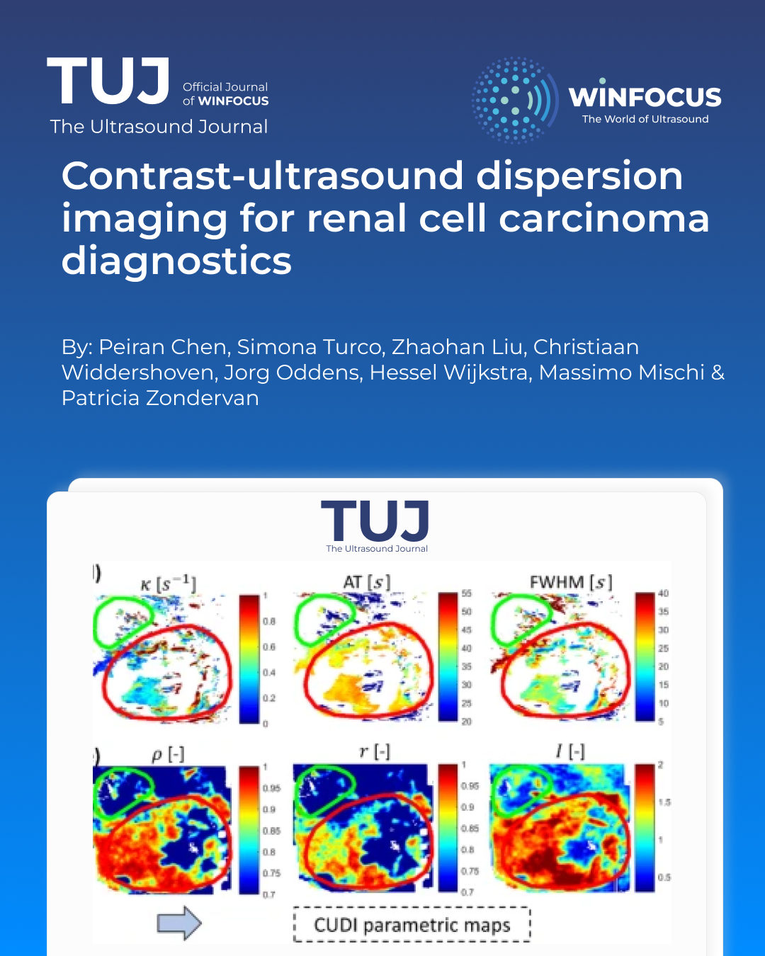
Cost-effective screening methods for Renal Cell Carcinoma (RCC) are still lacking. Angiogenesis is a recognized hallmark of cancer growth, leading to distinguishable perfusion patterns in tumors from those in normal tissue. This establishes the basis for diagnostic imaging solutions by dynamic contrast-enhanced ultrasound (DCE-US). In the past years, we have developed contrast-ultrasound dispersion imaging (CUDI) techniques to quantify prostate DCE-US acquisitions, obtaining promising results for prostate cancer localization. In this pilot study, we investigated for the first time its feasibility for RCC localization. DCE-US acquisitions of the kidney in 5 patients were used to perform CUDI analysis. With the obtained CUDI parameters and the delineated tumor and parenchyma regions, we performed pixel-based classification, from which the highest area under the receiver-operating-characteristic curve (AUC) = 0.96 was obtained for an individual patient, and an average AUC = 0.68 was obtained for the full patient dataset, showing the potential of CUDI for solid RCC localization. Further validation in a larger dataset and evaluation of the compatibility of point-of-care diagnosis are required.
References
1. Sung H, Ferlay J, Siegel RL et al (2021) Global cancer statistics 2020: GLOBOCAN estimates of incidence and mortality worldwide for 36 cancers in 185 countries. CA Cancer J Clin 71:209–249. https://doi.org/10.3322/caac.21660
2. Thurnher M, Putz T, Rahm A et al (2008) Renal cell carcinoma. Handb Dendritic Cells 3:1117–1127. https://doi.org/10.1002/9783527619696.ch53
3. Ljungberg B, Albiges L, Abu-Ghanem Y et al (2019) European association of urology guidelines on renal cell carcinoma: the 2019 update. Eur Urol 75:799–810. https://doi.org/10.1016/j.eururo.2019.02.011
4. Tosaka A, Ohya K, Yamada K et al (1990) Incidence and properties of renal masses and asymptomatic renal cell carcinoma detected by abdominal ultrasonography. J Urol 144:1097–1099. https://doi.org/10.1016/S0022-5347(17)39667-2
5. Usher-Smith J, Simmons RK, Rossi SH, Stewart GD (2020) Current evidence on screening for renal cancer. Nat Rev Urol 2020 1711 17:637–642. https://doi.org/10.1038/s41585-020-0363-3
6. FOLKMAN J (2002) Role of angiogenesis in tumor growth and metastasis. Semin Oncol 29:15–18. https://doi.org/10.1016/S0093-7754(02)70065-1
7. Weidner N, Carroll PR, Flax J et al (1993) Tumor angiogenesis correlates with metastasis in invasive prostate carcinoma. Am J Pathol 143:401
8. Russo G, Mischi M, Scheepens W et al (2012) Angiogenesis in prostate cancer: onset, progression and imaging. BJU Int 110:E794–E808. https://doi.org/10.1111/J.1464-410X.2012.11444.X
9. Miles KA, Lee TY, Goh V et al (2012) Current status and guidelines for the assessment of tumour vascular support with dynamic contrast-enhanced computed tomography. Eur Radiol 22:1430–1441. https://doi.org/10.1007/S00330-012-2379-4/TABLES/7
10. Sboros V, Tang MX (2010) The assessment of microvascular flow and tissue perfusion using ultrasound imaging. Proc Inst Mech Eng H 224:273–290. https://doi.org/10.1243/09544119JEIM621
11. Mao Y, Mu J, Zhao J et al (2018) The value of Superb microvascular imaging in differentiating benign renal mass from malignant renal tumor: A retrospective study. Br J Radiol 91. https://doi.org/10.1259/bjr.20170601
12. Harvey CJ, Alsafi A, Kuzmich S et al (2015) Role of US contrast agents in the assessment of indeterminate solid and cystic lesions in native and transplant kidneys. 35(1419–1432). https://doi.org/10.1148/rg2015140222
13. Xu ZF, Xu HX, Xie XY et al (2010) Renal cell carcinoma and renal angiomyolipoma: differential diagnosis with real-time contrast-enhanced ultrasonography. J Ultrasound Med 29:709–717. https://doi.org/10.7863/JUM.2010.29.5.709
14. Rübenthaler J, Negrão de Figueiredo G, Mueller-Peltzer K et al (2018) Evaluation of renal lesions using contrast-enhanced ultrasound (CEUS); a 10-year retrospective European single-centre analysis. Eur Radiol 28:4542–4549. https://doi.org/10.1007/S00330-018-5504-1
15. Ignee A, Straub B, Schuessler G, Dietrich CF (2010) Contrast enhanced ultrasound of renal masses. World J Radiol 2:15–31. https://doi.org/10.4329/wjr.v2.i1.15
16. Sun D, Wei C, Li Y et al (2016) Contrast-Enhanced ultrasonography with quantitative analysis allows differentiation of renal tumor histotypes. Sci Rep 2016 61 6:1–7. https://doi.org/10.1038/srep35081
17. Li CX, Lu Q, Huang BJ et al (2016) Quantitative evaluation of contrast-enhanced ultrasound for differentiation of renal cell carcinoma subtypes and Angiomyolipoma. Eur J Radiol 85:795–802. https://doi.org/10.1016/j.ejrad.2016.01.009
18. Delorme S, Knopp MV (1998) Non-invasive vascular imaging: assessing tumour vascularity. Eur Radiol 1998 84 8:517–527. https://doi.org/10.1007/S003300050428
19. Mischi M, Kuenen MPJ, Wijkstra H (2012) Angiogenesis imaging by Spatiotemporal analysis of ultrasound contrast agent dispersion kinetics. IEEE Trans Ultrason Ferroelectr Freq Control 59:621–629. https://doi.org/10.1109/TUFFC.2012.2241
20. Kuenen MPJ, Mischi M, Wijkstra H (2011) Contrast-ultrasound diffusion imaging for localization of prostate cancer. IEEE Trans Med Imaging 30:1493–1502. https://doi.org/10.1109/TMI.2011.2125981
21. Kuenen MPJ, Saidov TA, Wijkstra H et al (2013) Spatiotemporal correlation of ultrasound contrast agent Dilution curves for angiogenesis localization by dispersion imaging. IEEE Trans Ultrason Ferroelectr Freq Control 60:2665–2669. https://doi.org/10.1109/TUFFC.2013.2865
22. Kuenen MPJ, Saidov TA, Wijkstra H, Mischi M (2013) Contrast-Ultrasound dispersion imaging for prostate cancer localization by improved Spatiotemporal similarity analysis. Ultrasound Med Biol 39:1631–1641. https://doi.org/10.1016/J.ULTRASMEDBIO.2013.03.004
23. Schalk SG, Demi L, Bouhouch N et al (2017) Contrast-Enhanced ultrasound angiogenesis imaging by mutual information analysis for prostate cancer localization. IEEE Trans Biomed Eng 64:661–670. https://doi.org/10.1109/TBME.2016.2571624
24. Mannaerts CK, Engelbrecht MRW, Postema AW et al (2020) Detection of clinically significant prostate cancer in biopsy-naïve men: direct comparison of systematic biopsy, multiparametric MRI- and contrast-ultrasound-dispersion imaging-targeted biopsy. BJU Int 126:481–493. https://doi.org/10.1111/BJU.15093
25. Wagner RF, Smith SW, Sandrik JM, Lopez H (1983) Statistics of speckle in ultrasound B-Scans. IEEE Trans Sonics Ultrason 30:156–163. https://doi.org/10.1109/T-SU.1983.31404
26. Ta CN, Eghtedari M, Mattrey RF et al (2014) 2-tier in-plane motion correction and out-of-plane motion filtering for contrast-enhanced ultrasound. Invest Radiol 49:707. https://doi.org/10.1097/RLI.0000000000000074
27. Demené C, Deffieux T, Pernot M et al (2015) Spatiotemporal clutter filtering of ultrafast ultrasound data highly increases doppler and fUltrasound sensitivity. IEEE Trans Med Imaging 34:2271–2285. https://doi.org/10.1109/TMI.2015.2428634
28. Sammali F, Kuijsters NPM, Huang Y et al (2019) Dedicated ultrasound speckle tracking for quantitative analysis of uterine motion outside pregnancy. IEEE Trans Ultrason Ferroelectr Freq Control 66:581–590. https://doi.org/10.1109/TUFFC.2018.2867098
29. Li J, Huang X, Wang L et al (2024) Role of Contrast-Enhanced ultrasound with the enhancement pattern and qualitative analysis for differentiating hypovascular solid renal lesions. Ultrasound Med Biol 50:295–303. https://doi.org/10.1016/j.ultrasmedbio.2023.11.002
30. Tufano A, Drudi FM, Angelini F et al (2022) Contrast-Enhanced ultrasound (CEUS) in the evaluation of renal masses with histopathological Validation—Results from a prospective Single-Center study. Diagnostics 12:1–8. https://doi.org/10.3390/diagnostics12051209
31. Xue LY, Lu Q, Huang BJ et al (2015) Papillary renal cell carcinoma and clear cell renal cell carcinoma: differentiation of distinct histological types with contrast – enhanced ultrasonography. Eur J Radiol 84:1849–1856. https://doi.org/10.1016/J.EJRAD.2015.06.017
32. Cosgrove D (2003) Angiogenesis imaging - Ultrasound. Br J Radiol 76. https://doi.org/10.1259/BJR/86364648
33. Wildeboer RR, Van Sloun RJG, Schalk SG et al (2018) Convective-Dispersion modeling in 3D Contrast-Ultrasound imaging for the localization of prostate cancer. IEEE Trans Med Imaging 37:2593–2602. https://doi.org/10.1109/TMI.2018.2843396
34. van Sloun RJG, Demi L, Schalk SG et al (2018) Contrast-enhanced ultrasound tractography for 3D vascular imaging of the prostate. Sci Rep 8:14640. https://doi.org/10.1038/s41598-018-32982-2
35. Errico C, Pierre J, Pezet S et al (2015) Ultrafast ultrasound localization microscopy for deep super-resolution vascular imaging. Nature 527:499–502. https://doi.org/10.1038/nature16066
36. Bodard S, Denis L, Hingot V et al (2023) Ultrasound localization microscopy of the human kidney allograft on a clinical ultrasound scanner. Kidney Int 103:930–935. https://doi.org/10.1016/j.kint.2023.01.027
37. Chabouh G, Denis L, Bodard S et al (2024) Whole organ volumetric sensing ultrasound localization microscopy for characterization of kidney structure. IEEE Trans Med Imaging. https://doi.org/10.1109/TMI.2024.3411669
38. Christensen-Jeffries K, Couture O, Dayton PA et al (2020) Super-resolution ultrasound imaging. Ultrasound Med Biol 46:865–891. https://doi.org/10.1016/j.ultrasmedbio.2019.11.013
39. Wildeboer RR, Postema AW, Demi L et al (2017) Multiparametric dynamic contrast-enhanced ultrasound imaging of prostate cancer. Eur Radiol 27:3226–3234. https://doi.org/10.1007/s00330-016-4693-8
40. Wildeboer RR, van Sloun RJG, Huang P et al (2019) 3-D Multi-parametric Contrast-Enhanced ultrasound for the prediction of prostate cancer. Ultrasound Med Biol 45:2713–2724. https://doi.org/10.1016/J.ULTRASMEDBIO.2019.05.017
41. Chen P, Turco S, Wang Y et al (2024) Can 3D multiparametric ultrasound imaging predict prostate biopsy outcome? Ultrasound Med Biol 50:1194–1202. https://doi.org/10.1016/J.ULTRASMEDBIO.2024.04.007/ASSET/6DDFED5C-4E05-4535-BEE6-4999423131BF/MAIN.ASSETS/GR4.JPG
Downloads
Published
Issue
Section
License
Copyright (c) 2025 Peiran Chen, Simona Turco, Zhaohan Liu, Christiaan Widdershoven, Jorg Oddens, Hessel Wijkstra, Massimo Mischi, Patricia Zondervan (Author)

This work is licensed under a Creative Commons Attribution-NonCommercial 4.0 International License.
This is an Open Access article distributed under the terms of the Creative Commons Attribution License (https://creativecommons.org/licenses/by-nc/4.0) which permits unrestricted use, distribution, and reproduction in any medium, provided the original work is properly cited.
Authors retain the copyright for their published work. No formal permission will be required to reproduce parts (tables or illustrations) of published papers, provided the source is quoted appropriately and reproduction has no commercial intent.
How to Cite


