
Evaluation of a modified venous excess ultrasound (VExUS) protocol for estimation of venous congestion: a cohort study
Abstract
Background: Understanding venous congestion is critical to the management of many illnesses, but assessing volume status can be challenging. The current gold standard for volume status assessment of right heart catheterization (RHC) is invasive, costly, and often unavailable. Venous Excess Ultrasound Score (VExUS) is a novel ultrasound protocol for to assessment of venous congestion using the inferior vena cava, hepatic, portal and renal veins. Though there is a much interest in the technique, the renal component of the exam is challenging to acquire. For this reason we aimed to see if a modified VExUS (mVExUS) excluding the kidney component performs similarly to traditional VExUS (tVExUS) for detecting elevated right atrial pressure (RAP) as measured by RHC.
Methods: A consecutive cohort of 95 patients undergoing RHC had VExUS exams before the procedure. Researchers compared the performance of tVExUS, mVExUS, and inferior vena cava (IVC) diameter in predicting RAP > 12 mmHg.
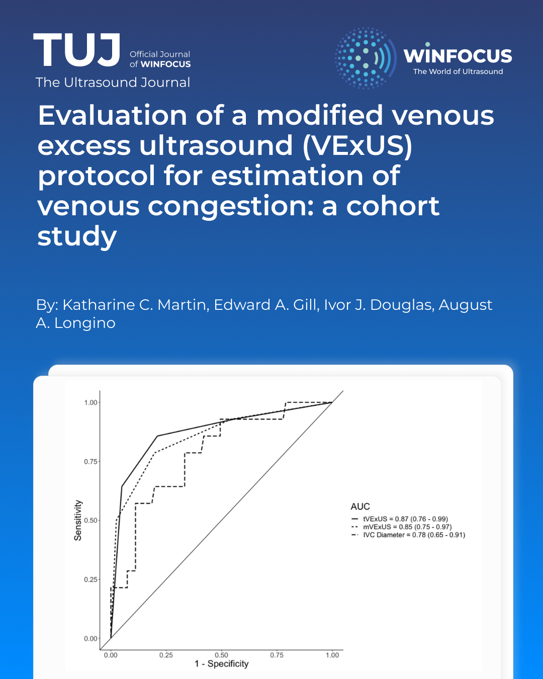
Results: The area under the curve (AUC) for detecting elevated RAP was similar for tVExUS (0.87) and mVExUS (0.85). Both methods achieved high sensitivity and specificity. Agreement between tVExUS and mVExUS scores was nearperfect (Cohen’s Kappa = 0.85).
Conclusion: mVExUS may be as effective as tVExUS in identifying elevated RAP. This abbreviated version could improve efficiency and adoption of VExUS for assessing venous congestion. Further studies are needed in diverse patient populations.
References
1. Beaubien-Souligny W, Benkreira A, Robillard P et al (2018) Alterations in portal vein flow and intrarenal venous flow are associated with acute kidney injury after cardiac surgery: a prospective observational cohort study. J Am Heart Assoc 7(19):e009961. https:// doi. org/ 10. 1161/ JAHA. 118. 009961
2. Boorsma EM, Ter Maaten JM, Damman K et al (2020) Congestion in heart failure: a contemporary look at physiology, diagnosis and treatment. Nat Rev Cardiol 17(10):641–655. https:// doi. org/ 10. 1038/ s41569- 020- 0379-7
3. Chen CY, Zhou Y, Wang P, Qi EY, Gu WJ (2020) Elevated central venous pressure is associated with increased mortality and acute kidney injury in critically ill patients: a meta-analysis. Crit Care 24(1):80. https:// doi. org/ 10. 1186/ s13054- 020- 2770-5
4. Chen KP, Cavender S, Lee J et al (2016) Peripheral edema, central venous pressure, and risk of AKI in critical illness. Clin J Am Soc Nephrol 11(4):602–608. https:// doi. org/ 10. 2215/ CJN. 08080 715
5. Li DK, Wang XT, Liu DW (2017) Association between elevated central venous pressure and outcomes in critically ill patients. Ann Intensive Care 7(1):83. https:// doi. org/ 10. 1186/ s13613- 017- 0306-1
6. Miller WL (2016) Fluid volume overload and congestion in heart failure: time to reconsider pathophysiology and how volume is assessed. Circ Heart Fail 9(8):e002922. https:// doi. org/ 10. 1161/ CIRCH EARTF AILURE. 115. 002922
7. Mullens W, Abrahams Z, Francis GS et al (2009) Importance of venous congestion for worsening of renal function in advanced decompensated heart failure. J Am Coll Cardiol 53(7):589–596. https:// doi. org/ 10. 1016/j. jacc. 2008. 05. 068
8. Banjade P, Subedi A, Ghamande S, Surani S, Sharma M (2023) Systemic venous congestion reviewed. Cureus. 15(8):e43716. https:// doi. org/ 10. 7759/ cureus. 43716
9. Koratala A, Ronco C, Kazory A (2022) Diagnosis of fluid overload: from conventional to contemporary concepts. Cardiorenal Med 12(4):141–154. https:// doi. org/ 10. 1159/ 00052 6902
10. Elhassan MG, Chao PW, Curiel A (2021) The conundrum of volume status assessment: revisiting current and future tools available for physicians at the bedside. Cureus. 13(5):e15253. https:// doi. org/ 10. 7759/ cureus. 15253
11. Breidthardt T, Moreno-Weidmann Z, Uthoff H et al (2018) How accurate is clinical assessment of neck veins in the estimation of central venous pressure in acute heart failure? Insights from a prospective study. Eur J Heart Fail 20(7):1160–1162. https:// doi. org/ 10. 1002/ ejhf. 1111
12. Hadian M, Pinsky MR (2006) Evidence-based review of the use of the pulmonary artery catheter: impact data and complications. Crit Care 10(Suppl 3):S8. https:// doi. org/ 10. 1186/ cc4834
13. Hoeper MM, Lee SH, Voswinckel R et al (2006) Complications of right heart catheterization procedures in patients with pulmonary hypertension in experienced centers. J Am Coll Cardiol 48(12):2546–2552. https:// doi. org/ 10. 1016/j. jacc. 2006. 07. 061
14. De Vecchis R, Baldi C, Giandomenico G, Di Maio M, Giasi A, Cioppa C (2016) Estimating right atrial pressure using ultrasounds: an old issue revisited with new methods. J Clin Med Res 8(8):569–574. https:// doi. org/ 10. 14740/ jocmr 2617w
15. Beaubien-Souligny W, Rola P, Haycock K et al (2020) Quantifying systemic congestion with Point-Of-Care ultrasound: development of the venous excess ultrasound grading system. Ultrasound J 12(1):16. https:// doi. org/ 10. 1186/ s13089- 020- 00163-w
16. Rola P, Miralles-Aguiar F, Argaiz E et al (2021) Clinical applications of the venous excess ultrasound (VExUS) score: conceptual review and case series. Ultrasound J 13(1):32. https:// doi. org/ 10. 1186/ s13089- 021- 00232-8
17. Jury D, Shaw AD (2021) Utility of bedside ultrasound derived hepatic and renal parenchymal flow patterns to guide management of acute kidney injury. Curr Opin Crit Care 27(6):587–592. https:// doi. org/ 10. 1097/ MCC. 00000 00000 000899
18. Gupta B, Ahluwalia P, Gupta A, Ranjan N, Kakkar K, Aneja P (2023) Utility of VExUS score in the peri-operative care unit, intensive care unit, and emergency setting—a systematic review. Indian J Anaesth 67(Suppl 4):S218–S226. https:// doi. org/ 10. 4103/ ija. ija_ 475_ 23
19. Longino A, Martin K, Leyba K et al (2023) Correlation between the VExUS score and right atrial pressure: a pilot prospective observational study. Crit Care 27(1):205. https:// doi. org/ 10. 1186/ s13054- 023- 04471-0
20. Prager R, Argaiz E, Pratte M et al (2023) Doppler identified venous congestion in septic shock: protocol for an international, multi-centre prospective cohort study (Andromeda-VEXUS). BMJ Open 13(7):e074843. https:// doi. org/ 10. 1136/ bmjop en- 2023- 074843
21. Rola P. The VExUS Course. Video. Accessed 2023. https:// vimeo. com/ ondem and/ theve xusco urse
22. Longino A, Martin K, Leyba K et al (2023) Prospective evaluation of venous excess ultrasound (VExUS) for estimation of venous congestion. Chest. https:// doi. org/ 10. 1016/j. chest. 2023. 09. 029
23. Brennan JM, Blair JE, Goonewardena S et al (2007) Reappraisal of the use of inferior vena cava for estimating right atrial pressure. J Am Soc Echocardiogr 20(7):857–861. https:// doi. org/ 10. 1016/j. echo. 2007. 01. 005
24. Patel AR, Alsheikh-Ali AA, Mukherjee J et al (2011) 3D echocardiography to evaluate right atrial pressure in acutely decompensated heart failure correlation with invasive hemodynamics. JACC Cardiovasc Imaging 4(9):938–945. https:// doi. org/ 10. 1016/j. jcmg. 2011. 05. 006
25. Prekker ME, Scott NL, Hart D, Sprenkle MD, Leatherman JW (2013) Pointof-care ultrasound to estimate central venous pressure: a comparison of three techniques. Crit Care Med 41(3):833–841. https:// doi. org/ 10. 1097/ CCM. 0b013 e3182 7466b7
Downloads
Published
Issue
Section
License
Copyright (c) 2025 Katharine C. Martin, Edward A. Gill, Ivor J. Douglas, August A. Longino (Author)

This work is licensed under a Creative Commons Attribution-NonCommercial 4.0 International License.
This is an Open Access article distributed under the terms of the Creative Commons Attribution License (https://creativecommons.org/licenses/by-nc/4.0) which permits unrestricted use, distribution, and reproduction in any medium, provided the original work is properly cited.
Authors retain the copyright for their published work. No formal permission will be required to reproduce parts (tables or illustrations) of published papers, provided the source is quoted appropriately and reproduction has no commercial intent.
How to Cite


