
Ultrasound evaluation of gallbladder wall thickness for predicting severe dengue: a systematic review and meta‑analysis
Keywords:
Dengue fever, Severe dengue, Gallbladder wall thickening (GBWT), Ultrasound, Risk predictionAbstract
Background: The prevalence of dengue fever (DF), a mosquito-borne viral disease, is rising worldwide. Its severe manifestations like thrombocytopenia and plasma leakage are associated with increased mortality. Ultrasound-detected gallbladder wall thickening (GBWT) has been suggested as a potential indicator of the severity of the disease.
Aims: This systematic review and meta-analysis evaluated the predictive value of GBWT in identifying patients at risk for severe dengue.
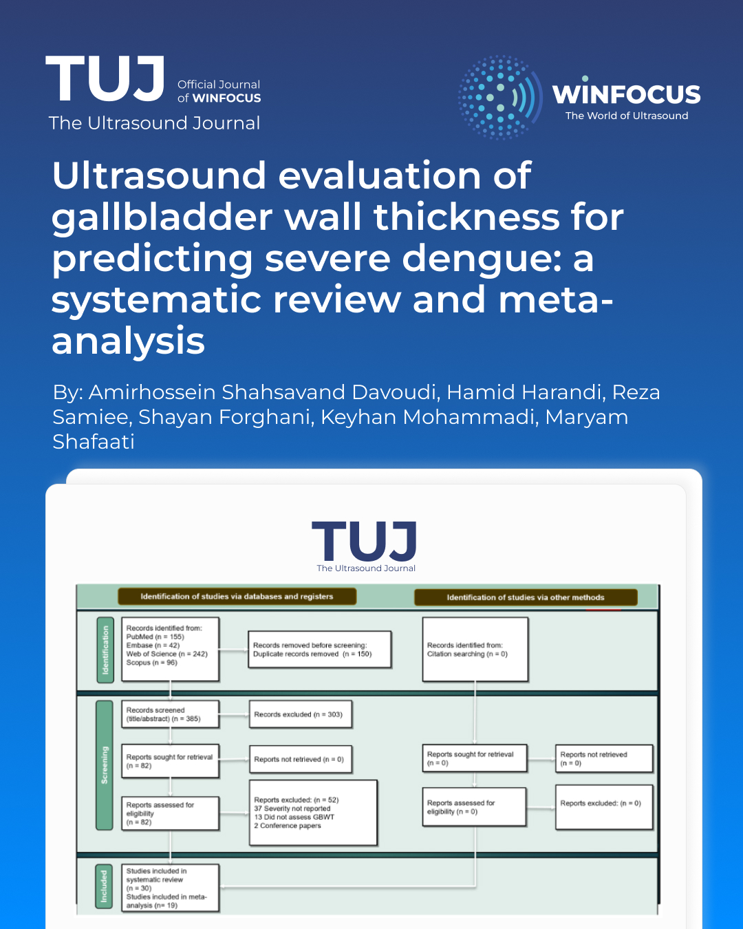
Methods: Following the PRISMA 2020 guidelines, we conducted a systematic search of Web of Science, PubMed, Embase, and Scopus. Among the inclusion criteria were original studies that assessed GBWT across various dengue severity categories. Then, we performed a meta-analysis using a random effects model and subgroup analyses based on severity criteria to determine the relationship between GBWT and severe dengue.
Results: For the meta-analysis, 19 studies qualified for the inclusion criteria. There was a significant association between GBWT and severe dengue, according to the odds ratio (OR) of 2.35 (95% CI 1.88–2.82, p < 0.001). The subgroup analysis revealed consistent results for thrombocytopenia (OR: 2.65) and plasma leakage (OR: 2.26), among other severity criteria.
Conclusions: A reliable ultrasound indicator, GBWT can help identify patients at risk for severe dengue early on, improving clinical decision-making and patient outcomes. However, the possibility of differential diagnosis requires cautious interpretation.
References
1. Lessa CLS, Hodel KVS, Gonçalves MDS, Machado BAS (2023) Dengue as a disease threatening global health: a narrative review focusing on Latin America and Brazil. Trop Med Infect Dis 8(5):241
2. Rigau-Pérez JG (2006) Severe dengue: the need for new case definitions. Lancet Infect Dis 6(5):297–302
3. World Health Organization. Dengue: guidelines for diagnosis, treatment, prevention and control: new edition. Available from: https://www.who.int/publications/i/item/9789241547871
4. Jaenisch T, Tam DTH, Kieu NTT, Van Ngoc T, Nam NT, Van Kinh N et al (2016) Clinical evaluation of dengue and identification of risk factors for severe disease: protocol for a multicentre study in 8 countries. BMC Infect Dis 16:1–11
5. Htun TP, Xiong Z, Pang J (2021) Clinical signs and symptoms associated with WHO severe dengue classification: a systematic review and meta-analysis. Emerg Microb Infect 10(1):1116–1128
6. World Health Organization (2010) Working to overcome the global impact of neglected tropical diseases. In: Crompton DWT (ed) First WHO report on neglected tropical diseases. WHO, Geneva
7. Soo K-M, Khalid B, Ching S-M, Tham CL, Basir R, Chee H-Y (2017) Meta-analysis of biomarkers for severe dengue infections. PeerJ 5:e3589
8. Yuan K, Chen Y, Zhong M, Lin Y, Liu L (2022) Risk and predictive factors for severe dengue infection: a systematic review and meta-analysis. PLoS ONE 17(4):e0267186
9. Shlaer WJ, Leopold GR, Scheible FW (1981) Sonography of the thickened gallbladder wall: a nonspecific finding. Am J Roentgenol 136(2):337–339
10. Setiawan MW, Samsi TK, Wulur H, Sugianto D, Pool TN (1998) Dengue haemorrhagic fever: ultrasound as an aid to predict the severity of the disease. Pediatr Radiol 28(1):1–4
11. Setiawan MW, Samsi TK, Pool TN, Sugianto D, Wulur H (1995) Gallbladder wall thickening in dengue hemorrhagic fever: an ultrasonographic study. J Clin Ultrasound 23(6):357–362
12. Santhosh VR, Patil PG, Srinath MG, Kumar A, Jain A, Archana M (2014) Sonography in the diagnosis and assessment of dengue fever. J Clin Imaging Sci. https://doi.org/10.4103/2156-7514.129260
13. Osorio L, Prieto I, Zuluaga D, Ropero D, Dewan N, Kirsch JD (2023) Evaluation of remote radiologist-interpreted point-of-care ultrasound for suspected dengue patients in a primary health care facility in Colombia. Infect Dis Poverty. https://doi.org/10.4103/2156-7514.129260
14. Page MJ, McKenzie JE, Bossuyt PM, Boutron I, Hoffmann TC, Mulrow CD et al (2021) The PRISMA 2020 statement: an updated guideline for reporting systematic reviews. BMJ 372:n71
15. Parmar JP, Mohan C, Vora M (2017) Patterns of gall bladder wall thickening in dengue fever: a mirror of the severity of disease. Ultrasound Int Open 3(02):E76–E81
16. Organization WH (1997) Dengue haemorrhagic fever: diagnosis, treatment, prevention and control. World Health Organization, Geneva
17. Newcastle-Ottawa Scale (NOS) Available from: https://www.ohri.ca/programs/clinical_epidemiology/nosgen.pdf
18. Modesti PA, Reboldi G, Cappuccio FP, Agyemang C, Remuzzi G, Rapi S et al (2016) Panethnic differences in blood pressure in Europe: a systematic review and meta-analysis. PLoS ONE 11(1):e0147601
19. Adil B, Rabbani A, Ahmed S, Arshad I Sr, Khalid MA (2020) Gall bladder wall thickening in dengue fever—aid in labelling dengue hemorrhagic fever and a marker of severity. Cureus 12(11):e11331
20. Agarwal N, Jain P (2016) Sonography in dengue fever: an adjunct to clinico-laboratory profile. Ind J Public Health Res Dev 7:299
21. Asghar MS, Yasmin F, Tahir MJ, Anwar S, Yaseen R, Yousaf Z (2022) Predictive analysis of gallbladder wall thickness as a marker for bleeding risk and need for transfusion in dengue patients. Jpn J Infect Dis 75(3):234–240
22. Bandyopadhyay D, Chattaraj S, Hajra A, Mukhopadhyay S, Ganesan V (2016) A study on spectrum of hepatobiliary dysfunctions and pattern of liver involvement in dengue infection. J Clin Diagn Res. https://doi.org/10.7860/JCDR/2016/16946.7784
23. Bharath Kumar Reddy KR, Laksmana RR, Veerappa BG, Shivananda S (2013) Ultrasonography as a tool in predicting the severity of dengue fever in children–a useful aid in a developing country. Pediatr Radiol 43(8):971–977
24. Chaudhary S, Manrai K, Dhagat P, Dudeja P, Sen D, Grewal DS et al (2023) Abdominal and chest ultrasonography: a predictor for disease progression in nonsevere dengue. Med J Armed Forces India 79(4):386–391
25. de Araújo TM, Pivoto João GA, Bastos MS, Lima Gimaque JB, Gomes Almeida AC, Ngo TT et al (2019) Clinical relevance of gallbladder wall thickening for dengue severity: a cross-sectional study. PLoS ONE. https://doi.org/10.1371/journal.pone.0218939
26. Donaldson CD, de Mel S, Clarice CSH, Thilakawardana BU, de Mel P, Shalindi M et al (2021) Admission ultrasonography as a predictive tool for thrombocytopenia and disease severity in dengue infection. Trans R Soc Trop Med Hyg 115(12):1396–1402
27. Sahana KS, Sujatha R (2015) Clinical profile of dengue among children according to revised who classification: analysis of a 2012 outbreak from Southern India. Indian J Pediatr 82(2):109–113
28. Tauseef A, Ijaz F, Chaudhary FA, Ali Z, Akram T, Aftab RK et al (2019) Role of interleukin-10 and abdominopelvic ultrasound as a potential predictor of disease severity in dengue hemorrhagic fever. Cureus J Med Sci. https://doi.org/10.7759/cureus.5249
29. Uthraraj NS, Sriraam LM, Hiriyur Prakash M, Kumar M, Palanisamy U, Chettiakkapalayam Venkatachalam KU (2022) Predictive factors for the complications of dengue fever in children: a retrospective analysis. Cureus 14(12):e33027
30. Vedaraju KS, Kumar KRV, Vijayaraghavachari TV (2016) Role of ultrasound in the assessment of dengue fever. Int J Sci Study 3(10):59 62
31. Sai PMV, Dev B, Krishnan R (2005) Role of ultrasound in dengue fever. Br J Radiol 78(929):416–418
32. Yousaf KR, Atiq S, Sheikh QS, Nisar MS, Mansoor Z, Khalid S (2011) Sonographic features of polyserositis as an adjunct to clinico-pathological parameters in diagnosing and predicting the severity of dengue fever. Pak J Med Health Sci 5(1):184–189
33. Zulkarnain I (2004) Gallbladder edema in dengue hemorrhagic fever and its association with haematocrit levels and type of infections. Acta Med Indones 36(2):84–86
34. Ibrahim MA, Hamzah SS, Md Noor J, Mohamad MIK, Mokhtar MF, Isa MR et al (2022) The association of ultrasound assessment of gallbladder wall thickness with dengue fever severity. Ultrasound J 14(1):13
35. Jain A, Vasnaik M, Singam A, Mudiganti V (2024) A Cross-sectional study on bedside abdominal ultrasound findings as a diagnostic and prognostic tool in dengue fever in manipal hospital, Bengaluru, India. Cureus 16(7):e64734
36. Mallhi TH, Khan AH, Adnan AS, Sarriff A, Khan YH, Jummaat F (2015) Clinico-laboratory spectrum of dengue viral infection and risk factors associated with dengue hemorrhagic fever: a retrospective study. BMC Infect Dis. https://doi.org/10.1186/s12879-015-1141-3
37. Michels M, Sumardi U, de Mast Q, Jusuf H, Puspita M, Dewi IMW et al (2013) The predictive diagnostic value of serial daily bedside ultrasonography for severe dengue in Indonesian adults. Plos Neglected Trop Dis. https://doi.org/10.1371/journal.pntd.0002277
38. Nainggolan L, Wiguna C, Hasan I, Dewiasty E (2018) Gallbladder wall thickening for early detection of plasma leakage in dengue infected adult patients. Acta Med Indones 50(3):193–199
39. Oliveira GA, Machado RC, Horvat JV, Gomes LE, Guerra LR, Vandesteen L et al (2010) Transient reticular gallbladder wall thickening in severe dengue fever: a reliable sign of plasma leakage. Pediatr Radiol 40(5):720–724
40. Parmar J, Vora M, Mohan C, Shah S, Mahajan H, Patel T (2019) Honeycomb” pattern of gallbladder wall thickening—a forward step in early diagnosis of “severe dengue fever. Ind J Radiol Imaging 29(01):14–18
41. Pothapregada S, Kullu P, Kamalakannan B, Thulasingam M (2016) Is ultrasound a useful tool to predict severe dengue infection? Indian J Pediatr 83(6):500–504
42. Binh PT, Matheus S, Huong VTQ, Deparis X, Marechal V (2009) Early clinical and biological features of severe clinical manifestations of dengue in Vietnamese adults. J Clin Virol 45(4):276–280
43. Chacko B, Subramanian G (2008) Clinical, laboratory and radiological parameters in children with dengue fever and predictive factors for dengue shock syndrome. J Trop Pediatr 54(2):137–140
44. Guo C, Zhou Z, Wen Z, Liu Y, Zeng C, Xiao D et al (2017) Global epidemiology of dengue outbreaks in 1990–2015: a systematic review and meta-analysis. Front Cell Infect Microbiol. https://doi.org/10.3389/fcimb.2017.00317
45. Byard RW (2016) Lethal dengue virus infection: a forensic overview. Am J Forensic Med Pathol 37(2):74–78
46. Tejo AM, Hamasaki DT, Menezes LM, Ho Y-L (2024) Severe dengue in the intensive care unit. J Intensive Med 4(1):16–33
47. Khan MB, Yang Z-S, Lin C-Y, Hsu M-C, Urbina AN, Assavalapsakul W et al (2023) Dengue overview: an updated systemic review. J Infect Public Health 16(10):1625–1642
48. Muller DA, Depelsenaire AC, Young PR (2017) Clinical and laboratory diagnosis of dengue virus infection. J Infect Dis 215(2):S89-s95
49. World Health Organization. Regional Office for South-East Asia. Comprehensive Guideline for Prevention and Control of Dengue and Dengue Haemorrhagic Fever. Revised and expanded edition.: WHO Regional Office for South-East Asia; 2011. Available from: https://iris.who.int/handle/10665/204894
50. Paraná VC, Feitosa CA, da Silva GCS, Gois LL, Santos LA (2024) Risk factors associated with severe dengue in Latin America: a systematic review and meta-analysis. Trop Med Int Health 29(3):173–191
51. Sangkaew S, Ming D, Boonyasiri A, Honeyford K, Kalayanarooj S, Yacoub S et al (2021) Risk predictors of progression to severe disease during the febrile phase of dengue: a systematic review and meta-analysis. Lancet Infect Dis 21(7):1014–1026
52. Chen CY, Chiu YY, Chen YC, Huang CH, Wang WH, Chen YH et al (2023) Obesity as a clinical predictor for severe manifestation of dengue: a systematic review and meta-analysis. BMC Infect Dis 23(1):502
53. Tsheten T, Clements ACA, Gray DJ, Adhikary RK, Furuya-Kanamori L, Wangdi K (2021) Clinical predictors of severe dengue: a systematic review and meta-analysis. Infect Dis Poverty 10(1):123
54. Moallemi S, Lloyd AR, Rodrigo C (2023) Early biomarkers for prediction of severe manifestations of dengue fever: a systematic review and a meta-analysis. Sci Rep 13(1):17485
55. Setiawan MW, Samsi TK, Wulur H, Sugianto D, Pool TN (1998) Dengue haemorrhagic fever: ultrasound as an aid to predict the severity of the disease. Pediatr Radiol 28:1–4
56. Md-Sani SS, Md-Noor J, Han W-H, Gan S-P, Rani N-S, Tan H-L et al (2018) Prediction of mortality in severe dengue cases. BMC Infect Dis 18(1):232
57. Talukdar S, Thanachartwet V, Desakorn V, Chamnanchanunt S, Sahassananda D, Vangveeravong M et al (2021) Predictors of plasma leakage among dengue patients in Thailand: a plasma-leak score analysis. PLoS ONE 16(7):e0255358
58. Srikiatkhachorn A (2009) Plasma leakage in dengue haemorrhagic fever. Thromb Haemost 102(6):1042–1049
59. Malavige GN, Ogg GS (2017) Pathogenesis of vascular leak in dengue virus infection. Immunology 151(3):261–269
60. Srikiatkhachorn A, Kelley JF (2014) Endothelial cells in dengue hemorrhagic fever. Antiviral Res 109(1):160–170
61. Runner GJ, Corwin MT, Siewert B, Eisenberg RL (2014) Gallbladder wall thickening. Am J Roentgenol 202(1):W1–W12
62. Davies P, Aoyagi Y (2017) Leptospirosis presenting as acute acalculous cholecystitis. Clin Case Rep 5(11):1775–1779
63. Curley JM, Mody RM, Gasser RA Jr (2011) Malaria caused by Plasmodium vivax complicated by acalculous cholecystitis. Am J Trop Med Hyg 85(1):42–49
64. Michels M, De Mast Q, Sumardi U, Van Der Ven AJ, Alisjahbana B (2012) Daily handheld ultrasonography performed by clinicians can detect subclinical plasma leakage and identify dengue patients at risk for dengue shock syndrome. Am J Trop Med Hyg 87(5):128
Downloads
Published
Issue
Section
License
Copyright (c) 2025 Amirhossein Shahsavand Davoudi, Hamid Harandi, Reza Samiee, Shayan Forghani, Keyhan Mohammadi, Maryam Shafaati (Author)

This work is licensed under a Creative Commons Attribution-NonCommercial 4.0 International License.
This is an Open Access article distributed under the terms of the Creative Commons Attribution License (https://creativecommons.org/licenses/by-nc/4.0) which permits unrestricted use, distribution, and reproduction in any medium, provided the original work is properly cited.
Authors retain the copyright for their published work. No formal permission will be required to reproduce parts (tables or illustrations) of published papers, provided the source is quoted appropriately and reproduction has no commercial intent.
How to Cite


