
Intraoperative elastography and spinal surgery: a systematic review of current and future applications in clinical and preclinical models
Keywords:
Ultrasound elastography, Spinal pathology, Intraoperative ultrasound, Surgical imagingAbstract
Background: While conventional imaging provides excellent structural detail of the spine, it cannot assess the mechanical properties of spinal tissue in real time. Ultrasound elastography (USE) is an emerging modality that quantifies tissue stiffness, offering a potential solution to this diagnostic gap. This review synthesizes the current evidence for the use of USE in spinal pathology.
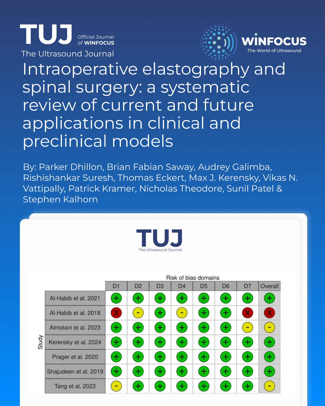
Main body: A systematic review of the PubMed, Cochrane, and Web of Science databases was conducted in accordance with PRISMA guidelines, yielding seven primary studies, three clinical and four preclinical, published between 2015 and 2024. These studies, comprising preclinical and clinical data, demonstrate USE's ability to provide real-time, quantitative feedback. Key applications identified include quantifying tension relief in tethered cord syndrome, differentiating spinal tumors from healthy tissue based on stiffness values, and assessing the biomechanical severity of acute and chronic spinal cord injury. Shear wave elastography (SWE) was the predominant modality, proving superior to strain elastography (SE) for spinal applications.
Conclusion: USE is a powerful adjunct to traditional spinal imaging, providing unique functional data that can enhance intraoperative surgical precision and decision-making. While challenges such as depth penetration and operator standardization remain, continued research and technological innovation position USE to significantly improve diagnostic accuracy and surgical outcomes in spinal disease management.
References
1. Ding W, Hu S, Wang P et al (2022) Spinal cord injury: the global incidence, prevalence, and disability from the global burden of disease study 2019. Spine 47(21):1532–1540. https://doi.org/10.1097/BRS.0000000000004417
2. Jannesar S, Salegio EA, Beattie MS, Bresnahan JC, Sparrey CJ (2021) Correlating tissue mechanics and spinal cord injury: patient-specific finite element models of unilateral cervical contusion spinal cord injury in non-human primates. J Neurotrauma 38(6):698–717. https://doi.org/10.1089/neu.2019.6840
3. Saway BF, Courtney J, Barley J, Frankel B, Hofstetter C, Kalhorn S (2024) Contrast enhanced ultrasound for traumatic spinal cord injury: an overview of current and future applications. Spinal Cord Ser Cases 10(1):31. https://doi.org/10.1038/s41394-024-00644-3
4. Albakr A, Ben-Israel D, Yang R et al (2023) Ultrasound elastography in neurosurgery: current applications and future perspectives. World Neurosurg 170:195-205.e1. https://doi.org/10.1016/j.wneu.2022.10.108
5. Chan SS, Colecchia A, Duarte RF, Bonifazi F, Ravaioli F, Bourhis JH (2020) Imaging in hepatic veno-occlusive disease/sinusoidal obstruction syndrome. Biol Blood Marrow Transplant 26(10):1770–1779. https://doi.org/10.1016/j.bbmt.2020.06.016
6. Taljanovic MS, Gimber LH, Becker GW et al (2017) Shear-wave elastography: basic physics and musculoskeletal applications. Radiographics 37(3):855–870. https://doi.org/10.1148/rg.2017160116
7. Hersh AM, Weber-Levine C, Jiang K et al (2022) Applications of elastography in operative neurosurgery: a systematic review. J Clin Neurosci 104:18–28. https://doi.org/10.1016/j.jocn.2022.07.019
8. Ganau M, Syrmos N, Martin AR, Jiang F, Fehlings MG (2018) Intraoperative ultrasound in spine surgery: history, current applications, future developments. Quant Imaging Med Surg 8(3):261–267. https://doi.org/10.21037/qims.2018.04.02
9. Hubertus V, Badhiwala JH, Hejrati N et al (2025) AO spine clinical practice recommendations for the surgical management of acute traumatic spinal cord injury: contemporary concepts. Glob Spine J 15(8):3572–3579. https://doi.org/10.1177/21925682251350941
10. Ali DM, Harrop J, Sharan A, Vaccaro AR, Sivaganesan A (2023) Technical aspects of intra-operative ultrasound for spinal cord injury and myelopathy: a practical review. World Neurosurg 170:206–218. https://doi.org/10.1016/j.wneu.2022.10.101
11. Tzschätzsch H, Kreft B, Schrank F, Bergs J, Braun J, Sack I (2018) In vivo time-harmonic ultrasound elastography of the human brain detects acute cerebral stiffness changes induced by intracranial pressure variations. Sci Rep 8(1):17888. https://doi.org/10.1038/s41598-018-36191-9
12. Sigrist RMS, Liau J, Kaffas AE, Chammas MC, Willmann JK (2017) Ultrasound elastography: review of techniques and clinical applications. Theranostics 7(5):1303–1329. https://doi.org/10.7150/thno.18650
13. Manbachi A (2022) The abundant promise of ultrasound in neurosurgery: a broad overview and thoughts on ethical paths to realizing its benefits, 1st edn. SPIE
14. Page MJ, McKenzie JE, Bossuyt PM et al (2021) The PRISMA 2020 statement: an updated guideline for reporting systematic reviews. Int J Surg 88:105906. https://doi.org/10.1016/j.ijsu.2021.105906
15. McGuinness LA, Higgins JPT (2021) Risk-of-bias VISualization (robvis): an R package and Shiny web app for visualizing risk-of-bias assessments. Res Synth Methods 12(1):55–61. https://doi.org/10.1002/jrsm.1411
16. Brezinski ME (2006) 9—Adjuvant techniques: absorption spectroscopy, contrast probes, phase contrast, elastography, and entangled photons. In: Brezinski ME (ed) Optical coherence tomography. Academic Press, pp 245–275b. https://doi.org/10.1016/B978-012133570-0/50011-1
17. Low G, Kruse SA, Lomas DJ (2016) General review of magnetic resonance elastography. World J Radiol 8(1):59–72. https://doi.org/10.4329/wjr.v8.i1.59
18. Altaf A, Baqai MWS, Urooj F et al (2023) Intraoperative use of ultra-low-field, portable magnetic resonance imaging—first report. Surg Neurol Int 14:212. https://doi.org/10.25259/SNI_124_2023
19. Al-Habib A, Alhothali W, Albakr A et al (2021) Effects of compressive lesions on intraoperative human spinal cord elasticity. J Neurosurg Spine 35(6):807–816. https://doi.org/10.3171/2021.1.SPINE201482
20. Kerensky MJ, Paul A, Routkevitch D et al (2024) Tethered spinal cord tension assessed via ultrasound elastography in computational and intraoperative human studies. Commun Med 4(1):4. https://doi.org/10.1038/s43856-023-00430-6
21. Judy BF, Kerensky M, Hersh A et al (2023) 668 intraoperative ultrasound elastography is a promising technology to quantify reduced spinal cord tension during posterior vertebral column subtraction osteotomy for recurrent tethered cord syndrome. Neurosurgery 69(Supplement_1):28. https://doi.org/10.1227/neu.0000000000002375_668
22. Almotairi FS, Basalamah AA, Amir A, Al-Habib AF (2023) Intraoperative demonstration of reduced distal spinal cord stiffness following untethering of the spinal cord using ultrasound shear wave elastography (SWE). World Neurosurgery: X 20:100225. https://doi.org/10.1016/j.wnsx.2023.100225
23. Venkatesh SK, Yin M, Glockner JF et al (2008) MR elastography of liver tumors: preliminary results. Am J Roentgenol 190(6):1534–1540. https://doi.org/10.2214/AJR.07.3123
24. Prager J, Adams CF, Delaney AM et al (2020) Stiffness-matched biomaterial implants for cell delivery: clinical, intraoperative ultrasound elastography provides a “target” stiffness for hydrogel synthesis in spinal cord injury. J Tissue Eng 11:2041731420934806. https://doi.org/10.1177/2041731420934806
25. Al-Habib A, Albakr A, Al Towim A et al (2018) In vivo assessment of spinal cord elasticity using shear wave ultrasound in dogs. J Neurosurg Spine 29(4):461–469. https://doi.org/10.3171/2018.2.SPINE171195
26. Shajudeen P, Tang S, Chaudhry A et al (2020) Modeling and analysis of ultrasound elastographic axial strains for spine fracture identification. IEEE Trans Ultrason Ferroelectr Freq Control 67(5):898–909. https://doi.org/10.1109/TUFFC.2019.2956730
27. Tang S, Weiner B, Taraballi F et al (2023) Assessment of spinal cord injury using ultrasound elastography in a rabbit model in vivo. Sci Rep 13(1):15323. https://doi.org/10.1038/s41598-023-41172-8
28. Ren ZX, Xu JH, Cheng X, Xu GX, Long HQ (2023) Pathophysiological mechanisms of chronic compressive spinal cord injury due to vascular events. Neural Regen Res 18(4):790. https://doi.org/10.4103/1673-5374.353485
29. Nouh MR (2019) Imaging of the spine: where do we stand? World J Radiol 11(4):55–61. https://doi.org/10.4329/wjr.v11.i4.55
30. Li J, Ma Y, Zhang T, Shung KK, Zhu B (2022) Recent advancements in ultrasound transducer: from material strategies to biomedical applications. BME Front. https://doi.org/10.34133/2022/9764501
31. Cui XW, Li KN, Yi AJ et al (2022) Ultrasound elastography. Endoscopic Ultrasound 11(4):252. https://doi.org/10.4103/EUS-D-21-00151
32. Huang R, Jiang L, Xu Y et al (2019) Comparative diagnostic accuracy of contrast-enhanced ultrasound and shear wave elastography in differentiating benign and malignant lesions: a network meta-analysis. Front Oncol 9:102. https://doi.org/10.3389/fonc.2019.00102
Downloads
Published
Issue
Section
License
Copyright (c) 2025 Parker Dhillon, Brian Fabian Saway, Audrey Galimba, Rishishankar Suresh, Thomas Eckert, Max J. Kerensky, Vikas N. Vattipally, Patrick Kramer, Nicholas Theodore, Sunil Patel, Stephen Kalhorn (Author)

This work is licensed under a Creative Commons Attribution-NonCommercial 4.0 International License.
This is an Open Access article distributed under the terms of the Creative Commons Attribution License (https://creativecommons.org/licenses/by-nc/4.0) which permits unrestricted use, distribution, and reproduction in any medium, provided the original work is properly cited.
Authors retain the copyright for their published work. No formal permission will be required to reproduce parts (tables or illustrations) of published papers, provided the source is quoted appropriately and reproduction has no commercial intent.
How to Cite


