
Ultrasound-based assessment of spinal muscle thickness and elasticity in patients with idiopathic scoliosis
Keywords:
Idiopathic scoliosis, Ultrasound, Muscle thickness, Muscle elasticityAbstract
Purpose: Utilizing ultrasonic imaging technology, this study assessed and compared the thickness and elasticity features of the abdominal and spinal back muscles in patients with idiopathic scoliosis to those of healthy individuals. The objective was to elucidate the mechanical adaptations in spinal muscles among IS patients.
Methods: This cross-sectional study included 38 patients diagnosed with idiopathic scoliosis and 33 healthy controls. Outcome measures comprised the Cobb angle, spinal curvature, muscle thickness, and muscle elasticity. Ultrasound elastography imaging was employed to assess the thickness and elasticity of the erector spinae, rectus abdominis, external oblique, and transverse abdominis muscles bilaterally at corresponding spinal levels. The objective was to document and compare the ultrasonic imaging characteristics of these muscles in individuals with idiopathic scoliosis and in the normal population.
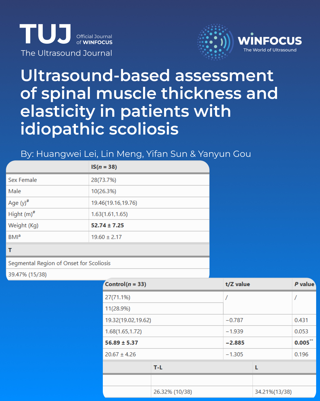
Results: The study findings indicated that idiopathic scoliosis patients had significantly lower body weight than the control group, with C7-CSVL notably greater in the idiopathic scoliosis group than in healthy individuals. Muscle thickness was substantially reduced on both the concave and convex sides at T6, T10, and L3 levels of the erector spinae, as well as in the rectus abdominis (RA) and transverse abdominis (TrA) muscles, relative to the normal cohort. Additionally, idiopathic scoliosis patients exhibited increased elasticity in the erector spinae muscle on the convex side at T6, while the elasticity of the erector spinae muscle on the concave side at L3 was significantly lower compared to healthy individuals.
Conclusions: This study, utilizing ultrasound elastography imaging technology, unveiled distinct features in individuals with mild idiopathic scoliosis, including decreased muscle thickness in the erector spinae at T6, T10, and L3 levels, as well as heightened elasticity in the thoracic region and reduced elasticity in the lumbar region. The findings presented in this study provide insights for diagnostic strategies in individuals with early-stage scoliosis.
References
1. Negrini S, Donzelli S, Aulisa AG, Czaprowski D, Schreiber S, de Mauroy JC, Diers H, Grivas TB, Knott P, Kotwicki T, Lebel A, Marti C, Maruyama T, O’Brien J, Price N, Parent E, Rigo M, Romano M, Stikeleather L, Wynne J, Zaina F (2018) 2016 SOSORT guidelines: orthopaedic and rehabilitation treatment of idiopathic scoliosis during growth. Scoliosis Spinal Disorders 13:3
2. Duncombe P, Izatt MT, Pivonka P, Claus A, Little JP, Tucker K (2023) Quantifying muscle size asymmetry in adolescent idiopathic scoliosis using Three-dimensional magnetic resonance imaging. Spine (Phila Pa 1976) 48(24):1717–1725
3. Chan WWY, Fu S, Chong T, Singh G, Tsai DSJ, Wong MCY, Zheng Y, Parent EC, Cheung JPY, Wong AYL (2024) Associations between paraspinal muscle characteristics and spinal curvature in conservatively treated adolescent idiopathic scoliosis: a systematic review. Spine Journal: Official J North Am Spine Soc 24(4):692–720
4. Ye H, Xu Y, Mi R, Liu Y, Lyu Y, Wu S, Wu G (2024) Evaluation of paravertebral muscle structure asymmetry in idiopathic scoliosis using imaging techniques. World Neurosurg 191:e547–e555
5. Wang Q, Li M, Lou EHM, Chu WCW, Lam T, Cheng JCY, Wong M (2016) Validity study of vertebral rotation measurement using 3-D ultrasound in adolescent idiopathic scoliosis. Ultrasound Med Biol 42(7):1473–1481
6. Zimmer M, Kleiser B, Marquetand J, Ateş F (2023) Shear wave elastography characterizes passive and active mechanical properties of biceps brachii muscle in vivo. J Mech Behav Biomed Mater 137:105543
7. Moreau B, Vergari C, Gad H, Sandoz B, Skalli W, Laporte S (2016) Non-invasive assessment of human multifidus muscle stiffness using ultrasound shear wave elastography: a feasibility study. Proc Inst Mech Eng H 230(8):809–814
8. Vergari C, Dubois G, Vialle R, Gennisson J, Tanter M, Dubousset J, Rouch P, Skalli W (2016) Lumbar annulus fibrosus Biomechanical characterization in healthy children by ultrasound shear wave elastography. Eur Radiol 26(4):1213–1217
9. Gharbi A, Obeid I, Larrieu D, Boissière L, Huneidi M, Lamotte-Paulet P, Tamir M, Aleman C, Charles YP (2024) Coronal alignment in normal individuals and moderate scoliosis: normative values, variation with age and comparison with sagittal alignment. Brain Spine 4:103917
10. Scaturro D, Balbo A, Vitagliani F, Stramazzo L, Camarda L, Letizia Mauro G (2022) Is there a Relationship between Idiopathic Scoliosis and Body Mass? A Scoping Review. LID – 10.3390/nu14194011 [doi] LID – 4011. Nutrients. 14(19)
11. Jeon K, Kim D (2018) The association between low body weight and scoliosis among Korean elementary school Students. 2018;15(12):2613. LID – 10.3390/ijerph15122613 [doi] LID – 2613. Int J Environ Res Public Health 15(12). https://doi.org/10.3390/ijerph15122613
12. Jeon K, Kim D (2021) Low body mass index levels and idiopathic scoliosis in Korean children: a Cross-Sectional Study. Children (Basel). 2021;8(7):570. LID – 10.3390/children8070570 [doi] LID – 570. Children (Basel, Switzerland) 8(7). https://doi.org/10.3390/children8070570
13. Yang H, Li Z, Hai Y, Zhang H (2023) The role of lumbosacral paraspinal muscle degeneration and low vertebral bone mineral density on distal instrumentation-related problems following long-instrumented spinal fusion for degenerative lumbar scoliosis: a retrospective cohort study. Quant Imaging Med Surg 13(7):4475–4492
14. Zhou M, Liu L, Chen Z, Ma B, Fu X, Cheng Y, Kan S, Liu C, Zhao X, Feng S, Jiang Z, Zhu R (2023) Characteristics of paraspinal muscle degeneration in patients with adult degenerative scoliosis. European spine journal: official publication of the European spine Society, the European spinal deformity Society, and the European section of the cervical. Spine Res Soc 32(11):4020–4029
15. Zapata KA, Wang-Price SS, Sucato DJ, Dempsey-Robertson M (2015) Ultrasonographic measurements of paraspinal muscle thickness in adolescent idiopathic scoliosis: a comparison and reliability study. Pediatr Phys Therapy: Official Publication Sect Pediatr Am Phys Therapy Association 27(2):119–125
16. Liu Y, Pan A, Hai Y, Li W, Yin L, Guo R (2019) Asymmetric Biomechanical characteristics of the paravertebral muscle in adolescent idiopathic scoliosis. Clin Biomech (Bristol Avon) 65:81–86
17. Modi HN, Suh S, Yang J, Hong J, Venkatesh K, Muzaffar N (2010) Spontaneous regression of curve in immature idiopathic scoliosis - does spinal column play a role to balance? An observation with literature review. J Orthop Surg Res 5:80
18. Millner PA, Dickson RA (1996) Idiopathic scoliosis: biomechanics and biology. Eur Spine J 5(6):362–373. https://doi.org/10.1007/BF00301963
19. Sima Borna PNPL (2017) Ultrasound measurements of the lateral abdominal muscle thicknesses in girls with adolescent idiopathic scoliosis. Asian J Sports Med 1(8):e32274
20. Hides J, Wilson S, Stanton W, Mcmahon S, Keto H, Mcmahon K, Bryant M, Richardson C (2006) An MRI investigation into the function of the transversus abdominis muscle during drawing-in of the abdominal wall. Spine (Phila Pa 1976) 31(6):E175–E178
21. Mcmeeken JM, Beith ID, Newham DJ, Milligan P, Critchley DJ (2004) The relationship between EMG and change in thickness of transversus abdominis. Clin Biomech (Bristol Avon) 19(4):337–342
22. Maughan RJ, Watson JS, Weir J (1983) Strength and cross-sectional area of human skeletal muscle. J Physiol 338:37–49
23. Gou Y, Tao J, Lei H, Hou M, Chen X, Wang X (2024) Trunk kinematic analysis of ascent and descent stairs in college students with idiopathic scoliosis: a case-control study. Spine Journal: Official J North Am Spine Soc 24(9):1712–1722
24. Driscoll M, Aubin C, Moreau A, Villemure I, Parent S (2009) The role of spinal concave-convex biases in the progression of idiopathic scoliosis. Eur Spine Journal: Official Publication the Eur Spine Soc the 18(2):180–187
25. Weng H, Li Q (2022) Effect of Core Stability Training on Correction and Surface Electronic Signals of Paravertebral in Adolescent Idiopathic Scoliosis. Biomed Res Int. 2022: 1819606
26. Ong RYL, Thazhakkattu Vasu D, Jun LK, Yuet NJ, Isaac Fernandez M, Selvakumar K, Ming Zi Goh J (2025) Effectiveness of dynamic neuromuscular stabilization approach in lumbopelvic stability and gait parameters in individuals with idiopathic scoliosis: a randomized controlled trial. Med (Baltim) 104(12):e41905
Downloads
Published
Issue
Section
License
Copyright (c) 2025 Huangwei Lei, Lin Meng, Yifan Sun, Yanyun Gou (Author)

This work is licensed under a Creative Commons Attribution-NonCommercial 4.0 International License.
This is an Open Access article distributed under the terms of the Creative Commons Attribution License (https://creativecommons.org/licenses/by-nc/4.0) which permits unrestricted use, distribution, and reproduction in any medium, provided the original work is properly cited.
Authors retain the copyright for their published work. No formal permission will be required to reproduce parts (tables or illustrations) of published papers, provided the source is quoted appropriately and reproduction has no commercial intent.
How to Cite


