
Cerebrovascular reactivity metrics as predictors of cognitive performance in healthy ageing: insights from transcranial colour‑coded ultrasound
Keywords:
Cerebrovascular reactivity, Physiologic, Transcranial colour-coded Doppler ultrasound, Breath-holding, hyperventilation, cognitive impairmentAbstract
Introduction: This study was designed to investigate the utility of cerebrovascular reactivity (CVR) metrics, derived from transcranial colour-coded Doppler ultrasound (TCCD). Three main CVR metrics were examined as potential markers for cerebrovascular risk associated with mild cognitive impairment (MCI), a stage between normal cognition and dementia.
Methods: We investigated 122 eligible, stroke-free, healthy, community-based Chinese adults (mean age, 65.34 ± 6.86 years). Cognitive performance was assessed using the validated Hong Kong version of the Montreal Cognitive Assessment. On a scale of 0–30, participants with low scores < 26 (modelled according to level of education) were designated to have a mild neurocognitive disorder or MCI. Following the measurement of cerebrovascular conductance (CVC) derived from cerebral blood flow and mean arterial pressure, three physiologic CVR metrics were assessed. The CVR assessments were based on restricted 30 s breath-holding, 60 s hyperventilation, and an unrestricted breath-holding index (BHI), respectively quantified using transcranial colour-coded Doppler ultrasound. The predictabilities and associations between CVR metrics, haemodynamic parameters, and cognitive performance were statistically investigated.
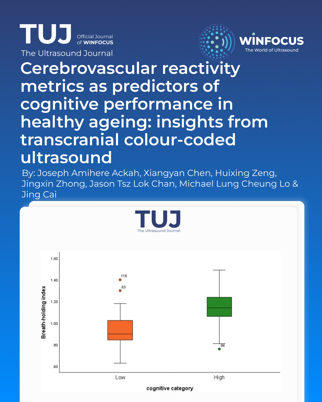
Results: Using TCCD, BHI emerged as the most accurate and robust metric of CVR for predicting mild cognitive disorders [AUC 0.827 (95% CI 0.725, 0.930)] and independently predicted overall cognitive performance, highlighting its clinical value for early identification of at-risk individuals. The three CVR metrics outperformed CVC in predicting mild cognitive impairment and were distinctively correlated. Although CVR measures by breath-holding and BHI were closely related (r = 0.704, 95% CI 0.598, 0.786, p < 0.001), Bland–Altman analysis revealed that they are not interchangeable, indicating the importance of metric selection for accurate cerebrovascular assessment.
Conclusion: The BHI, derived from simple and clinically tolerable methods, demonstrates clear potential to enhance the prediction and early identification of vascular cognitive impairment in healthy adults. By leveraging insights from cerebral haemodynamics, TCCD-based cerebrovascular risk screening may enable more effective and targeted interventions, ultimately contributing to better long-term cognitive health outcomes.
References
1. Staszewski J, Skrobowska E, Piusińska-Macoch R, Brodacki B, Stępień A (2019) Cerebral and extracerebral vasoreactivity in patients with different clinical manifestations of cerebral small-vessel disease: data from the significance of hemodynamic and hemostatic factors in the course of different manifestations of cerebral small-ves. J Ultrasound Med 38:975–987
2. Sforza M, Bianchini E, Alivernini D, Salvetti M, Pontieri FE, Sette G (2022) The impact of cerebral vasomotor reactivity on cerebrovascular diseases and cognitive impairment. J Neural Transm 129:1321–1330
3. Shim Y, Yoon B, Shim DS, Kim W, An JY, Yang DW (2015) Cognitive correlates of cerebral vasoreactivity on transcranial Doppler in older adults. J Stroke Cerebrovasc Dis 24:1262–1269
4. Miller KB, Howery AJ, Rivera-Rivera LA, Johnson SC, Rowley HA, Wieben O et al (2019) Age-related reductions in cerebrovascular reactivity using 4D flow MRI. Front Aging Neurosci 11:1–11
5. Daher A, Payne S (2024) The conducted vascular response as a mediator of hypercapnic cerebrovascular reactivity: a modelling study. Comput Biol Med 170:107985
6. Regenhardt RW, Nolan NM, Das AS, Mahajan R, Monk AD, LaRose SL et al (2024) Transcranial Doppler cerebrovascular reactivity: thresholds for clinical significance in cerebrovascular disease. J Neuroimaging 34:348–355
7. Blair GW, Thrippleton MJ, Shi Y, Hamilton I, Stringer M, Chappell F et al (2020) Intracranial hemodynamic relationships in patients with cerebral small vessel disease. Neurology 94:E2258–E2269
8. Bishop CCR, Powell S, Rutt D, Browse NL (1986) Transcranial doppler measurement of middle cerebral artery blood flow velocity: a validation study. Stroke 17:913–915
9. Takahashi S, Tanizaki Y, Kimura H, Akaji K, Kano T, Suzuki K et al (2015) Prediction of cerebrovascular reserve capacity by computed tomography perfusion using 320-row computed tomography. J Stroke Cerebrovasc Dis 24:939–945
10. Boas DA, Gaudette T, Strangman G, Cheng X, Marota JJA, Mandeville JB (2001) The accuracy of near infrared spectroscopy and imaging during focal changes in cerebral hemodynamics. Neuroimage 13:76–90
11. Huo C, Xu G, Li W, Xie H, Zhang T, Liu Y et al (2021) A review on functional near-infrared spectroscopy and application in stroke rehabilitation. Med Nov Technol Devices 11:100064
12. Fisher JA, Venkatraghavan L, Mikulis DJ (2018) Magnetic resonance imaging-based cerebrovascular reactivity and hemodynamic reserve a review of method optimization and data interpretation. Stroke 49:2011–2018
13. Gunda ST, Ng TKV, Liu TY, Chen Z, Han X, Chen X et al (2024) A comparative study of Transcranial Color-Coded Doppler (TCCD) and Transcranial Doppler (TCD) ultrasonography techniques in assessing the intracranial cerebral arteries haemodynamics. Diagnostics. https://doi.org/10.3390/diagnostics14040387
14. Kargiotis O, Safouris A, Magoufis G, Georgala M, Roussopoulou A, Stamboulis E et al (2018) The role of neurosonology in the diagnosis and management of patients with carotid artery disease: a review. J Neuroimaging 28:239–251
15. Rudziński W, Swiat M, Tomaszewski M, Krejza J (2007) Cerebral hemodynamics and investigations of cerebral blood flow regulation. Nucl Med Rev 10:29–42
16. Claassen JAHR, Zhang R, Fu Q, Witkowski S, Levine BD (2007) Transcranial Doppler estimation of cerebral blood flow and cerebrovascular conductance during modified rebreathing. J Appl Physiol 102:870–877
17. Staszewski J, Dȩbiec A, Skrobowska E, Stȩpień A (2021) Cerebral vasoreactivity changes over time in patients with different clinical manifestations of cerebral small vessel disease. Front Aging Neurosci 13:1–12
18. Wolf ME (2014) Functional TCD: regulation of cerebral hemodynamics—cerebral autoregulation, vasomotor reactivity, and neurovascular coupling. Front Neurol Neurosci 36:40–56
19. Settakis G, Lengyel A, Molnár C, Bereczki D, Csiba L, Fülesdi B (2002) Transcranial Doppler study of the cerebral hemodynamic changes during breath-holding and hyperventilation tests. J Neuroimaging 12:252–258
20. Markus HS, Harrison MJG (1992) Estimation of cerebrovascular reactivity using transcranial Doppler, including the use of breath-holding as the vasodilatory stimulus. Stroke 23:668–673
21. Bian Y, Wang JC, Sun F, Sun ZY, Lin YJ, Liu Y et al (2019) Assessment of cerebrovascular reserve impairment using the breath-holding index in patients with leukoaraiosis. Neural Regen Res 14:1412–1418
22. Panerai RB, Brassard P, Burma JS, Castro P, Claassen JAHR, van Lieshout JJ et al (2023) Transfer function analysis of dynamic cerebral autoregulation: a CARNet white paper 2022 update. J Cereb Blood Flow Metab 43:3–25
23. Kostoglou K, Bello-Robles F, Brassard P, Chacon M, Claassen JAH, Czosnyka M et al (2024) Time-domain methods for quantifying dynamic cerebral blood flow autoregulation: review and recommendations. A white paper from the Cerebrovascular Research Network (CARNet). J Cereb Blood Flow Metab. https://doi.org/10.1177/0271678X241249276/FORMAT/EPUB
24. Wei EP, Kontos HA, Patterson JL (1980) Dependence of pial arteriolar response to hypercapnia on vessel size. Am J Physiol 238:697–703. https://doi.org/10.1152/ajpheart19802385H697
25. Akazawa N, Kumagai H, Yoshikawa T, Myoenzono K, Tanahashi K, Maeda S (2021) Cerebral blood flow velocity is associated with endothelial function in men. J Mens Health 17:41–46
26. Ide K, Eliasziw M, Poulin MJ (2003) Relationship between middle cerebral artery blood velocity and end-tidal PCO2 in the hypocapnic-hypercapnic range in humans. J Appl Physiol 95:129–137
27. Lin W, Xiong L, Han J, Leung T, Leung H, Chen X et al (2014) Hemodynamic effect of external counterpulsation is a different measure of impaired cerebral autoregulation from vasoreactivity to breath-holding. Eur J Neurol 21:326–331
28. Nowakowska-Kotas M, Nowakowska-Kotas M, Lisiak W, Wiśniewska E, Budrewicz S, Wiśniewski P (2025) Breath-holding index—a new approach to an old method. Eur J Transl Clin Med 8:10–18
29. Lim EY, Yang DW, Cho AH, Shim YS (2018) Cerebrovascular hemodynamics on transcranial Doppler ultrasonography and cognitive decline in mild cognitive impairment. J Alzheimers Dis 65:651–657
30. Bhandari A, Feridooni T, Pikula A, Styra R, Mikulis DJ, Howe KL (2024) Evaluating the influence of altered cerebral hemodynamics on cognitive performance in asymptomatic carotid artery stenosis: a systematic review. J Vasc Surg 79:436–447
31. Murrell CJ, Cotter JD, Thomas KN, Lucas SJE, Williams MJA, Ainslie PN (2013) Cerebral blood flow and cerebrovascular reactivity at rest and during sub-maximal exercise: effect of age and 12 week exercise training. Age (Omaha) 35:905–920
32. Silvestrini M, Pasqualetti P, Baruffaldi R, Bartolini M, Handouk Y, Matteis M et al (2006) Cerebrovascular reactivity and cognitive decline in patients with Alzheimer disease. Stroke 37:1010–1015
33. Pinto J, Bright MG, Bulte DP, Figueiredo P (2021) Cerebrovascular reactivity mapping without gas challenges: a methodological guide. Front Physiol 11:1–19
34. Moore CL, Henry DS, McClenahan SJ, Ball KK, Rusch NJ, Rhee SW (2021) Metoprolol impairs β1-adrenergic receptor-mediated vasodilation in rat cerebral arteries: implications for β-blocker therapy. J Pharmacol Exp Ther 376:127–135
35. Cseplo P, Vamos Z, Ivic I, Torok O, Toth A, Koller A et al (2016) The beta-1-receptor blocker nebivolol elicits dilation of cerebral arteries by reducing smooth muscle i. journals.plos.org. PLoS ONE 2016(11):e0164010
36. Erdfelder E, Faul F, Buchner A, Lang AG (2009) Statistical power analyses using G*power 3.1: tests for correlation and regression analyses. Behav Res Methods 41:1149–1160
37. Kang H (2021) Sample size determination and power analysis using the G*power software. J Educ Eval Health Prof 18:1–12
38. Wong A, Xiong YY, Kwan PWL, Chan AYY, Lam WWM, Wang K et al (2009) The validity, reliability and clinical utility of the Hong Kong montreal cognitive assessment (HK-MoCA) in patients with cerebral small vessel disease. Dement Geriatr Cogn Disord 28:81–87
39. Chan MYM, Ling YT, Chen XY, Chan ST, Kwong KK, Zheng YP (2023) Success rate of transcranial Doppler scanning of cerebral arteries at different transtemporal windows in healthy elderly individuals. Ultrasound Med Biol 49:588–598
40. Li Y, Li F, Li Y, Cui X, Li J, Zhi H et al (2020) Effect of cuff positioning on the accuracy of blood pressure measurement with automated electronic blood pressure monitors. J Clin Hypertens 22:1163–1172
41. Muntner P, Shimbo D, Carey RM, Charleston JB, Gaillard T, Misra S et al (2019) Measurement of blood pressure in humans: a scientific statement from the American heart association. Hypertension 73:E35–E66
42. Fagan TC, Conrad KA, Mayshar PV, Mackie MJ, Hagaman RM (1988) Single versus triplicate measurements of blood pressure and heart rate. Hypertension 11:282–284
43. Lasek-Bal A, Kazibutowska Z, Gołba A, Motta E (2012) Cerebral vasoreactivity in hypocapnia and hypercapnia in patients with diabetes mellitus type 2 with or without arterial hypertension. Neurol Neurochir Pol 46:529–535
44. Ochs MM, Kajzar I, Salatzki J, Ochs AT, Riffel J, Osman N et al (2021) Hyperventilation/breath-hold maneuver to detect myocardial ischemia by strain-encoded CMR: diagnostic accuracy of a needle-free stress protocol. JACC Cardiovasc Imaging 14:1932–1944
45. Koo TK, Li MY (2016) A guideline of selecting and reporting intraclass correlation coefficients for reliability research. J Chiropr Med 15:155
Downloads
Published
Issue
Section
License
Copyright (c) 2025 Joseph Amihere Ackah, Xiangyan Chen, Huixing Zeng, Jingxin Zhong, Jason Tsz Lok Chan, Michael Lung Cheung Lo, Jing Cai (Author)

This work is licensed under a Creative Commons Attribution-NonCommercial 4.0 International License.
This is an Open Access article distributed under the terms of the Creative Commons Attribution License (https://creativecommons.org/licenses/by-nc/4.0) which permits unrestricted use, distribution, and reproduction in any medium, provided the original work is properly cited.
Authors retain the copyright for their published work. No formal permission will be required to reproduce parts (tables or illustrations) of published papers, provided the source is quoted appropriately and reproduction has no commercial intent.
How to Cite


