
Lung ultrasound assessment of pulmonary effects of large patent ductus arteriosus in extremely preterm infants beyond the transitional period
Keywords:
Neonates, Lung ultrasound, Echocardiography, Patent ductus arteriosus, Preterm infantAbstract
Background: Several studies have suggested a positive association between elevated lung ultrasound scores (LUS) and large patent ductus arteriosus (L-PDA), although findings remain inconsistent. Lung ultrasound score, a semi-quantitative measure of pulmonary aeration loss, has been proposed as a surrogate marker of excessive lung fluid, which may reflect the hemodynamic burden of a significant PDA. The aim of this study was to assess the association between LUS and L-PDA in preterm neonates beyond the initial transitional period and examine its correlations with echocardiographic measures of ductal shunting. This is a cohort retrospective study that included preterm infants born at < 29 weeks’ gestation who underwent LUS within 24 h of targeted neonatal echocardiography. Infants were categorized as having L-PDA (diameter ≥ 1.5 mm, left-to-right shunt) or no/small PDA (< 1.5 mm). Clinical characteristics, LUS, and echocardiographic parameters including PDA diameter, left atrial-to-aortic root (LA: Ao) ratio, and left ventricular output (LVO) were compared. Statistical analyses included univariate, multivariate, and correlation assessments.
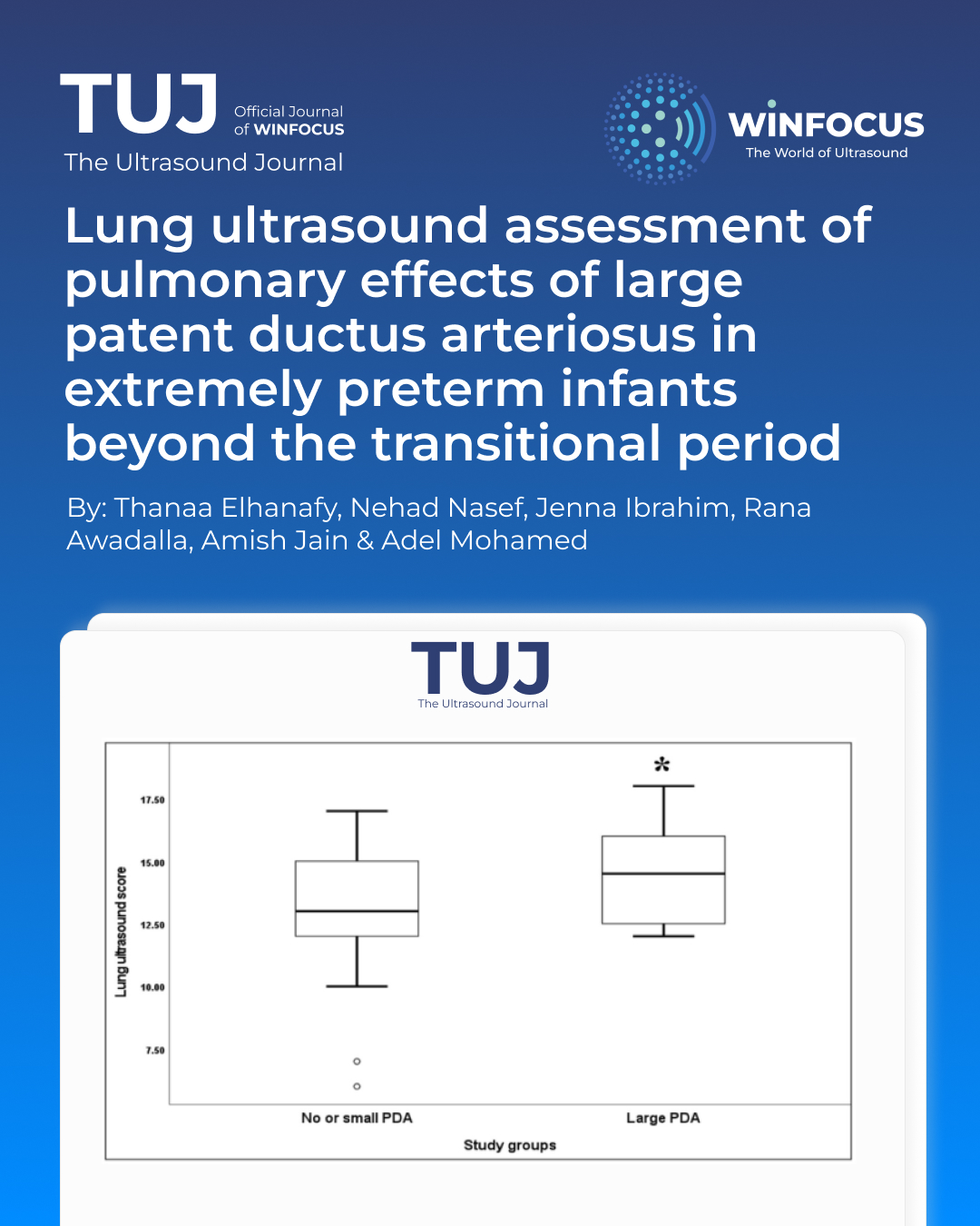
Results: Among 119 infants included in the analysis, 56 (47%) had L-PDA, and 63 (53%) had no or small PDA. Infants with L-PDA had significantly lower gestational age and higher rates of invasive ventilation. LUS, LA: Ao ratio, and LVO were significantly elevated in the L-PDA group (all p < 0.001). LUS correlated with PDA diameter (r = 0.27, p = 0.003) and respiratory severity score (r = 0.49, p < 0.001). Furthermore, LUS was found to be independently predictive for L-PDA (adjusted OR 1.5; 95% CI: 1.1–1.9). Each 1-point increase in LUS was associated with a 0.14 mm increase in PDA diameter. Inter-rater reliability for LUS was strong (IRR = 0.86).
Conclusion: Beyond the transitional period, LUS was significantly associated with PDA size and independently predicted L-PDA in extremely preterm infants.
References
1. Sellmer A, Bjerre JV, Schmidt MR, McNamara PJ, Hjortdal VE, Host B, Bech BH, Henriksen TB (2013) Morbidity and mortality in preterm neonates with patent ductus arteriosus on day 3. Arch Dis Child Fetal Neonatal Ed 98(6):F505–510
2. Clyman RI (2018) Patent ductus arteriosus, its treatments, and the risks of pulmonary morbidity. Semin Perinatol 42(4):235–242
3. Hansmann G, Sallmon H, Roehr CC, Kourembanas S, Austin ED, Koestenberger M (2021) European pediatric pulmonary vascular disease N: pulmonary hypertension in bronchopulmonary dysplasia. Pediatr Res 89(3):446–455
4. Schena F, Francescato G, Cappelleri A, Picciolli I, Mayer A, Mosca F, Fumagalli M (2015) Association between hemodynamically significant patent ductus arteriosus and bronchopulmonary dysplasia. J Pediatr 166(6):1488–1492
5. Mirza H, Garcia J, McKinley G, Hubbard L, Sensing W, Schneider J, Oh W, Wadhawan R (2019) Duration of significant patent ductus arteriosus and bronchopulmonary dysplasia in extremely preterm infants. J Perinatol 39(12):1648–1655
6. Jain A, Shah PS (2015) Diagnosis, evaluation, and management of patent ductus arteriosus in preterm neonates. JAMA Pediatr 169(9):863–872
7. Mohamed A, Mohsen N, Ibrahim J, Lee S, Kharrat A, Shah PS, Jain A (2025) Association of lung ultrasound score with large patent ductus arteriosus in preterm neonates during the transitional period. Eur J Pediatr 184(6):348
8. Skelton R, Evans N, Smythe J (1994) A blinded comparison of clinical and echocardiographic evaluation of the preterm infant for patent ductus arteriosus. J Paediatr Child Health 30(5):406–411
9. Martini S, Gatelli IF, Vitelli O, Vitali F, De Rienzo F, Parladori R, Corvaglia L, Martinelli S (2023) Impact of patent ductus arteriosus on non-invasive assessments of lung fluids in very preterm infants during the transitional period. Eur J Pediatr 182(9):4247–4251
10. Zong H, Huang Z, Lin B, Zhao J, Fu Y, Yu Y, Sun H, Yang C (2023) The predictive value of lung ultrasound score on hemodynamically significant patent ductus arteriosus among neonates. Diagnostics (Basel) 13(13)
11. Savoia M, McNamara PJ, Titolo A, Cattarossi L (2022) Lung ultrasound score parallels trends in systemic haemodynamics after PDA ligation: a case series. Eur J Pediatr 181(6):2541–2546
12. Zhao M, Huang XM, Niu L, Ni WX, Zhang ZQ (2020) Lung ultrasound score predicts the extravascular lung water content in Low-Birth-Weight neonates with patent ductus arteriosus. Med Sci Monit 26:e921671
13. Louis D, Belen K, Farooqui M, Idiong N, Amer R, Hussain A, ElSayed Y (2021) Prone versus supine position for lung ultrasound in neonates with respiratory distress. Am J Perinatol 38(2):176–181
14. Brat R, Yousef N, Klifa R, Reynaud S, Shankar Aguilera S, De Luca D (2015) Lung ultrasonography score to evaluate oxygenation and surfactant need in neonates treated with continuous positive airway pressure. JAMA Pediatr 169(8):e151797
15. Jain A, El-Khuffash AF, Kuipers BCW, Mohamed A, Connelly KA, McNamara PJ, Jankov RP, Mertens L (2017) Left ventricular function in healthy term neonates during the transitional period. J Pediatr 182:197–203e192
16. McNamara PJ, Jain A, El-Khuffash A, Giesinger R, Weisz D, Freud L, Levy PT, Bhombal S, de Boode W, Leone T et al (2024) Guidelines and recommendations for targeted neonatal echocardiography and cardiac Point-of-Care ultrasound in the neonatal intensive care unit: an update from the American society of echocardiography. J Am Soc Echocardiogr 37(2):171–215
17. Mitra S, McNamara PJ (2020) Patent ductus Arteriosus-Time for a definitive trial. Clin Perinatol 47(3):617–639
18. Mitra S, Florez ID, Tamayo ME, Mbuagbaw L, Vanniyasingam T, Veroniki AA, Zea AM, Zhang Y, Sadeghirad B, Thabane L (2018) Association of placebo, indomethacin, ibuprofen, and acetaminophen with closure of hemodynamically significant patent ductus arteriosus in preterm infants: A systematic review and Meta-analysis. JAMA 319(12):1221–1238
19. McNamara PJ, Sehgal A (2007) Towards rational management of the patent ductus arteriosus: the need for disease staging. Arch Dis Child Fetal Neonatal Ed 92(6):F424–427
20. El-Khuffash A, James AT, Corcoran JD, Dicker P, Franklin O, Elsayed YN, Ting JY, Sehgal A, Malikiwi A, Harabor A et al (2015) A patent ductus arteriosus severity score predicts chronic lung disease or death before discharge. J Pediatr 167(6):1354–1361 e1352
21. Singh Y, Tissot C, Fraga MV, Yousef N, Cortes RG, Lopez J, Sanchez-de-Toledo J, Brierley J, Colunga JM, Raffaj D et al (2020) International evidence-based guidelines on point of care ultrasound (POCUS) for critically ill neonates and children issued by the POCUS working group of the European society of paediatric and neonatal intensive care (ESPNIC). Crit Care 24(1):65
22. Ozdemir M, Tepe T, Ozlu F, Yapicioglu H, Atmis A, Demir F, Unal I, Narli N (2024) Lung ultrasound score in the decision of patent ductus arteriosus closure in neonates. J Clin Ultrasound 52(4):415–425
23. Noori S, McCoy M, Friedlich P, Bright B, Gottipati V, Seri I, Sekar K (2009) Failure of ductus arteriosus closure is associated with increased mortality in preterm infants. Pediatrics 123(1):e138–144
24. El-Khuffash AF, McNamara PJ (2011) The patent ductus arteriosus ligation decision. J Pediatr 158(6):1037–1038 author reply 1038–1039
25. Khositseth A, Nuntnarumit P, Chongkongkiat P (2011) Echocardiographic parameters of patent ductus arteriosus in preterm infants. Indian Pediatr 48(10):773–778
26. Raimondi F, Migliaro F, Corsini I, Meneghin F, Dolce P, Pierri L, Perri A, Aversa S, Nobile S, Lama S et al (2021) Lung ultrasound score progress in neonatal respiratory distress syndrome. Pediatrics 147(4)
Downloads
Published
Issue
Section
License
Copyright (c) 2025 Thanaa Elhanafy, Nehad Nasef, Jenna Ibrahim, Rana Awadalla, Amish Jain, Adel Mohamed (Author)

This work is licensed under a Creative Commons Attribution-NonCommercial 4.0 International License.
This is an Open Access article distributed under the terms of the Creative Commons Attribution License (https://creativecommons.org/licenses/by-nc/4.0) which permits unrestricted use, distribution, and reproduction in any medium, provided the original work is properly cited.
Authors retain the copyright for their published work. No formal permission will be required to reproduce parts (tables or illustrations) of published papers, provided the source is quoted appropriately and reproduction has no commercial intent.
How to Cite


