
Noninvasive assessment of hemodynamic profile and myocardial mechanics in pulsus alternans patients by multiple echocardiographic methods
Keywords:
Pulsus alternans, Left ventricular, Systolic,, Echocardiography, StrainAbstract
Background: Pulsus alternans (PA) is an intriguing phenomenon and a clinically rare entity. Accurately assessing cardiac function in patients with PA remains challenging. This study aims to investigate the myocardial mechanical characteristics and non-invasive hemodynamic profiles of PA patients using multiple echocardiographic imaging modalities.
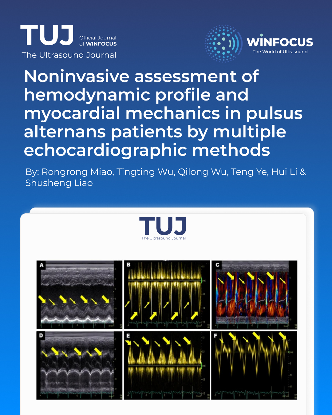
Methods: Clinical and echocardiographic data were retrospectively analysed from 16 patients diagnosed with PA by echocardiography at our hospital between January 2021 and May 2025. In this study, the characteristics of PA were elaborated by multiple echocardiographic methods, and the non-invasive hemodynamic profile was determined by pulse-wave Doppler.
Results: Sixteen patients were enrolled. Seven were classified as NYHA class III and six as class IV. Elevated levels of NT-proBNP and hs-cTNT were observed in most patients. Follow-up ranged from 1 to 44 months, and five patients experienced adverse outcomes, including heart transplantation, rehospitalisation, and death. Within this cohort, three patients exhibited biventricular PA, while 13 patients presented with left ventricular (LV) PA. Key hemodynamic parameters varied significantly: LVOT-VTIstrong beat ranged from 11.3 cm to 29.2 cm, LVOT-VTIweak beat from 6.8 cm to 22.1 cm, and the variation rate between strong and weak beats (∆LVOT-VTI) ranged from 19 to 52%. Global longitudinal strain (GLS) was significantly reduced in 14 patients (range: − 1.2% to − 10.4%), while peak strain dispersion (PSD) increased (range: 47 ms to 117.5 ms). Two patients were excluded from strain analysis due to suboptimal imaging. Hemodynamic parameters (LVOT-VTIstrong beat, LVOT-VTIweak beat and ∆LVOT-VTI) showed strong correlations with GLS in PA patients (r = 0.806, P = 0.001; r = 0.642, P = 0.018 and r = 0.611, P = 0.027, respectively). NT-proBNP was significantly positively related to adverse outcomes in PA patients (r = 0.669, P = 0.012).
Conclusion: Echocardiography is essential for evaluating cardiac function in patients with PA. This study used multiple echocardiographic methods to delineate the characteristics of this intriguing clinical phenomenon. Non-invasive hemodynamic parameters are potentially important for prognosis assessment, and myocardial strain assessment provides valuable insights into myocardial mechanical features. A comprehensive analysis using multimodality imaging is crucial for accurately identifying this disease, potentially enhancing the understanding of the pathophysiological mechanism of PA.
References
1. Traube L, Fall von E (1872) Pulsus Bigeminus nebst Bemerkungen uberdie Leberschwellungen bei Klappenfehlern and uber acute Leberatrophie [in German]. Berlin Klin Wochenschr 9:185–188
2. Surawicz B, Fisch C (1992) Cardiac alternans: diverse mechanisms and clinical manifestations. J Am Coll Cardiol 20(2):483–499. https://doi.org/10.1016/0735-1097(92)90122-4
3. Nguyen T, Cao LB, Tran M et al (2013) Biventricular pulsus alternans: an echocardiographic finding in patient with pulmonary embolism. World J Clin Cases 1(5):162–165. https://doi.org/10.12998/wjcc.v1.i5.162
4. Perk G, Tunick PA, Kronzon I (2007) Systolic and diastolic pulsus alternans in severe heart failure. J Am Soc Echocardiogr 20(7):905e5–905e7. https://doi.org/10.1016/j.echo.2006.12.004
5. Madanat L, Jabri A, Bloomingdale R et al (2024) Concomitant systolic and diastolic doppler alternans: an ominous sign of left ventricular dysfunction. Cureus 8(10):16. https://doi.org/10.7759/cureus.71074
6. Aslanger E, Aggül B, Albayrak DG (2025) Pulsus alternans: caught in action. Catheter Cardiovasc Interv 105(1):270–271. https://doi.org/10.1002/ccd.31295
7. Cournand A, Ferrer MI, Harvey RM et al (1956) Cardiocirculatory studies in pulsus alternans of the systemic and pulmonary circulations. Circulation 14(2):163–174. https://doi.org/10.1161/01.cir.14.2.163
8. Michaels AD, Browne AE, Varghese P et al (2000) Intracoronary measurement of pulsus alternans. Catheter Cardiovasc Interv 51(3):335–338.
9. Kang Y, Yu L, Zhang Q (2020) Biventricular pulsus alternans: stolen pulses seen by echocardiography in pulmonary hypertension. Am J Med Sci 359(4):247–248. https://doi.org/10.1016/j.amjms.2019.09.013
10. Lab MJ, Seed WA (1993) Pulsus alternans. Cardiovasc Res 27(8):1407–1412. https://doi.org/10.1093/cvr/27.8.1407
11. Eisner DA, Caldwell JL, Kistamás K et al (2017) Calcium and Excitation-Contraction coupling in the heart. Circ Res 121(2):181–195. https://doi.org/10.1161/CIRCRESAHA.117.310230
12. Cooper T, Braunwald E, Morrow AG (1958) Pulsus alternans in aortic stenosis; hemodynamic observations in 50 patients studied by left heart catheterization. Circulation 18(1):64–70. https://doi.org/10.1161/01.cir.18.1.64
13. Weiss JN, Karma A, Shiferaw Y et al (2006) From pulsus to pulseless: the Saga of cardiac alternans. Circ Res 98(10):1244–1253. https://doi.org/10.1161/01.RES.0000224540.97431.f0
14. Voigt JU, Pedrizzetti G, Lysyansky P et al (2015) Definitions for a common standard for 2D speckle tracking echocardiography: consensus document of the eacvi/ase/industry task force to standardize deformation imaging. Eur Heart J Cardiovasc Imaging 16(1):1–11. https://doi.org/10.1093/ehjci/jeu184
15. Smiseth OA, Rider O, Cvijic M et al (2025) Myocardial strain imaging: theory, current practice, and the future. JACC Cardiovasc Imaging 18(3):340–381. https://doi.org/10.1016/j.jcmg.2024.07.011
16. Dini FL, Barletta V, Ballo P et al (2025) Left ventricular outflow indices in chronic systolic heart failure: thresholds and prognostic value. Echocardiography 42(2):e70109. https://doi.org/10.1111/echo.70109
17. NobleS, Ibrahim R (2009) Pulsus alternans in critical aortic stenosis. Can J Cardiol 25(7):e268. https://doi.org/10.1016/s0828-282x(09)70522-7
18. Saffitz JE, Corradi D (2016) The electrical heart: 25 years of discovery in cardiac electrophysiology, arrhythmias and sudden death. Cardiovasc Pathol 25(2):149–157. https://doi.org/10.1016/j.carpath.2015.11.005
19. Wilson AJ, Sands GB, LeGrice IJ et al (2022) Myocardial mesostructure and mesofunction. Am J Physiol Heart Circ Physiol 323(2):H257–H275. https://doi.org/10.1152/ajpheart.00059.2022
20. Castiglione V, Aimo A, Vergaro G et al (2022) Biomarkers for the diagnosis and management of heart failure. Heart Fail Rev 27(2):625–643. https://doi.org/10.1007/s10741-021-10105-w
21. Kuwahara K (2021) The natriuretic peptide system in heart failure: diagnostic and therapeutic implications. Pharmacol Ther 227:107863. https://doi.org/10.1016/j.pharmthera.2021.10786
Downloads
Published
Issue
Section
License
Copyright (c) 2025 Rongrong Miao, Tingting Wu, Qilong Wu, Teng Ye, Hui Li, Shusheng Liao (Author)

This work is licensed under a Creative Commons Attribution-NonCommercial 4.0 International License.
This is an Open Access article distributed under the terms of the Creative Commons Attribution License (https://creativecommons.org/licenses/by-nc/4.0) which permits unrestricted use, distribution, and reproduction in any medium, provided the original work is properly cited.
Authors retain the copyright for their published work. No formal permission will be required to reproduce parts (tables or illustrations) of published papers, provided the source is quoted appropriately and reproduction has no commercial intent.
How to Cite


