
WINFOCUS worldwide survey on central venous catheter insertion and position confirmation practices (CVC‑ICON study)
Keywords:
Patient safety, Hospital resources, Radiation exposure, Low-resource settingsAbstract
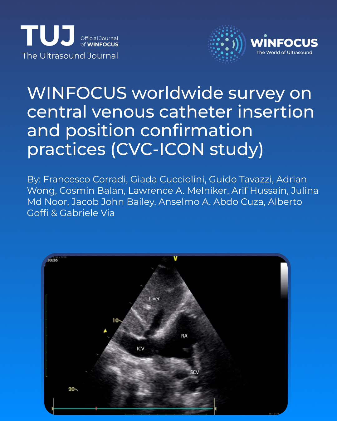
Background: Central venous catheters (CVC) are essential in medicine for monitoring, drug and fluid administration, and renal replacement therapy. Complications such as arrhythmias, endothelial damage, thrombosis, or hemothorax might arise from incorrect positioning. Despite evidence showing their reduction using ultrasound to guide insertion and correct tip positioning, and greater accuracy for tip position assessment vs. chest-X-ray (CXR), ultrasound adoption greatly varies worldwide. This study, conducted by the World Interactive Network Focused On Critical Ultrasound (WINFOCUS) aimed to assess global practices in CVC insertion and tip position confirmation.
Methods: A web-based survey was conducted (April–September 2023) among WINFOCUS members/affiliates across five continents. It assessed clinical backgrounds, CVC insertion and tip position check methods, and reasons for not using ultrasound. Developed by WINFOCUS Research sub-committee, the survey was emailed, with two reminders. Data were analyzed using SPSS 27.0.
Results: A total of 1,227 respondents (5.1% response rate) participated, mainly from Europe (33.5%), Asia (28.3%), and the Americas (30.9%), with 95.4% being physicians. Over half (51.3%) had over six years of experience and placed over 200 CVC, mostly using ultrasound guidance (70% of cases). The internal jugular vein (IJV) was the preferred insertion site (74%). Ultrasound was used for pre-insertion assessment (55%) and vessel puncture (57%) but less for guidewire confirmation (44%). CXR remained the primary method for tip position assessment (52%), while only 12% relied solely on bedside ultrasound. Barriers to exclusive ultrasound use included institutional guidelines (33.9%) and medico-legal concerns (13.8%).
Conclusions: Despite evidence favoring ultrasound for CVC insertion and tip position confirmation, its use remains inconsistent, with CXR still widely used. This survey underscores the need for standardized protocols and training to enhance US adoption, improve patient safety, and reduce CXR reliance.
References
1. McGee DC, Gould MK (2003) Preventing complications of central venous catheterization. N Engl J Med 348:1123–1133. https://doi.org/10.1056/NEJMra011883
2. Fletcher SJ, Bodenham AR (2000) Safe placement of central venous catheters: Where should the tip of the catheter lie? Br J Anaesth 85:188–191. https://doi.org/10.1093/bja/85.2.188
3. Raad II, Luna M, Khalil SA, Costerton JW, Lam C, Bodey GP (1994) The relationship between the thrombotic and infectious complications of central venous catheters. JAMA 271:1014–1016
4. Timsit JF, Farkas JC, Boyer JM, Martin JB, Misset B, Renaud B, Carlet J (1998) Central vein catheter-related thrombosis in intensive care patients: incidence, risks factors, and relationship with catheter-related sepsis. Chest 114:207–213. https://doi.org/10.1378/chest.114.1.207
5. Lamperti M, Biasucci DG, Disma N, Pittiruti M, Breschan C, Vailati D, Subert M, Traškaitė V, Macas A, Estebe J-P, Fuzier R, Boselli E, Hopkins P (2020) European Society of Anaesthesiology guidelines on peri-operative use of ultrasound-guided for vascular access (PERSEUS vascular access). Eur J Anaesthesiol 37:344–376. https://doi.org/10.1097/EJA.0000000000001180
6. Practice Guidelines for Central Venous Access (2020) An updated report by the american society of anesthesiologists task force on central venous access. Anesthesiology 132:8–43. https://doi.org/10.1097/ALN.0000000000002864
7. Corradi F, Guarracino F, Santori G, Brusasco C, Tavazzi G, Via G, Mongodi S, Mojoli F, Biagini RUD, Isirdi A, Dazzi F, Robba C, Vetrugno L, Forfori F, UCARE research group, (2022) Ultrasound localization of central vein catheter tip by contrast-enhanced transthoracic ultrasonography: a comparison study with trans-esophageal echocardiography. Crit Care 26:113. https://doi.org/10.1186/s13054-022-03985-3
8. Maizel J, Bastide M-A, Richecoeur J, Frenoy E, Lemaire C, Sauneuf B, Dupont H, Tamion F, Nseir S, Du Cheyron D, BoReal Study group (2016) Practice of ultrasound-guided central venous catheter technique by the French intensivists: a survey from the BoReal study group. Ann Intensive Care 6:76. https://doi.org/10.1186/s13613-016-0177-
9. Soni NJ, Reyes LF, Keyt H, Arango A, Gelfond JA, Peters JI, Levine SM, Adams SG, Restrepo MI (2016) Use of ultrasound guidance for central venous catheterization: a national survey of intensivists and hospitalists. J Crit Care 36:277–283. https://doi.org/10.1016/j.jcrc.2016.07.014
10. Burns KEA, Duffett M, Kho ME, Meade MO, Adhikari NKJ, Sinuff T, Cook DJ, Accademy Group (2008) A guide for the design and conduct of self-administered surveys of clinicians. CMAJ 179:245–252. https://doi.org/10.1503/cmaj.080372
11. Massicotte P, South A (2024) rnaturalearth: World Map Data from Natural Earth
12. R Core Team (2023) R: a language and environment for statistical computing
13. Wickham H (2016) ggplot2: elegant graphics for data analysis
14. Schmidt GA, Blaivas M, Conrad SA, Corradi F, Koenig S, Lamperti M, Saugel B, Schummer W, Slama M (2019) Ultrasound-guided vascular access in critical illness. Intensive Care Med 45:434–446. https://doi.org/10.1007/s00134-019-05564-7
15. Aslamy Z, Dewald CL, Heffner JE (1998) MRI of central venous anatomy: implications for central venous catheter insertion. Chest 114:820–826. https://doi.org/10.1378/chest.114.3.820
16. Reynolds N, McCulloch AS, Pennington CR, MacFadyen RJ (2001) Assessment of distal tip position of long-term central venous feeding catheters using transesophageal echocardiology. JPEN J Parenter Enteral Nutr 25:39–41. https://doi.org/10.1177/014860710102500139
17. Vezzani A, Brusasco C, Corradi F (2014) Contrast-enhanced ultrasound to determine correct central venous catheter position. Am J Emerg Med 32:809–810. https://doi.org/10.1016/j.ajem.2014.01.033
18. Meggiolaro M, Scatto A, Zorzi A, Roman-Pognuz E, Lauro A, Passarella C, Bonaccorso G (2015) Confirmation of correct central venous catheter position in the preoperative setting by echocardiographic “bubble-test.” Minerva Anestesiol 81:989–1000
19. Gidaro A, Casella F, Lugli F, Cogliati C, Calloni M, Samartin F, Brena N, Pace G (2023) Contrast enhanced ultrasound as a new tool to estimate the performance of midline catheters in the single patient. J Vasc Access 24:284–288. https://doi.org/10.1177/11297298211034629
20. Bou Chebl R, Kiblawi S, El Khuri C, El Hajj N, Bachir R, Aoun R, Abou Dagher G (2017) Use of contrast-enhanced ultrasound for confirmation of central venous catheter placement: systematic review and meta-analysis. J Ultrasound Med 36:2503–2510. https://doi.org/10.1002/jum.14296
21. Jauss M, Zanette E (2000) Detection of right-to-left shunt with ultrasound contrast agent and transcranial doppler sonography. Cerebrovasc Dis 10:490–496. https://doi.org/10.1159/000016119
22. Fan S, Nagai T, Luo H, Atar S, Naqvi T, Birnbaum Y, Lee S, Siegel RJ (1999) Superiority of the combination of blood and agitated saline for routine contrast enhancement. J Am Soc Echocardiogr 12:94–98. https://doi.org/10.1016/s0894-7317(99)70120-3
23. Jeon D-S, Luo H, Iwami T, Miyamoto T, Brasch AV, Mirocha J, Naqvi TZ, Siegel RJ (2002) The usefulness of a 10% air-10% blood-80% saline mixture for contrast echocardiography: doppler measurement of pulmonary artery systolic pressure. J Am Coll Cardiol 39:124–129. https://doi.org/10.1016/s0735-1097(01)01698-9
24. Timsit J-F, Baleine J, Bernard L, Calvino-Gunther S, Darmon M, Dellamonica J, Desruennes E, Leone M, Lepape A, Leroy O, Lucet J-C, Merchaoui Z, Mimoz O, Misset B, Parienti J-J, Quenot J-P, Roch A, Schmidt M, Slama M, Souweine B, Zahar J-R, Zingg W, Bodet-Contentin L, Maxime V (2020) Expert consensus-based clinical practice guidelines management of intravascular catheters in the intensive care unit. Ann Intensive Care 10:118. https://doi.org/10.1186/s13613-020-00713-4
25. Vezzani A, Manca T, Brusasco C, Santori G, Cantadori L, Ramelli A, Gonzi G, Nicolini F, Gherli T, Corradi F (2017) A randomized clinical trial of ultrasound-guided infra-clavicular cannulation of the subclavian vein in cardiac surgical patients: short-axis versus long-axis approach. Intensive Care Med 43:1594–1601. https://doi.org/10.1007/s00134-017-4756-6
26. Ablordeppey EA, Drewry AM, Theodoro DL, Tian L, Fuller BM, Griffey RT (2019) Current practices in central venous catheter position confirmation by point of care ultrasound: a survey of early adopters. Shock 51:613–618. https://doi.org/10.1097/SHK.0000000000001218
Downloads
Published
Issue
Section
License
Copyright (c) 2025 Francesco Corradi, Giada Cucciolini, Guido Tavazzi, Adrian Wong, Cosmin Balan, Lawrence A. Melniker, Arif Hussain, Julina Md Noor, Jacob John Bailey, Anselmo A. Abdo Cuza, Alberto Goffi, Gabriele Via (Author)

This work is licensed under a Creative Commons Attribution-NonCommercial 4.0 International License.
This is an Open Access article distributed under the terms of the Creative Commons Attribution License (https://creativecommons.org/licenses/by-nc/4.0) which permits unrestricted use, distribution, and reproduction in any medium, provided the original work is properly cited.
Authors retain the copyright for their published work. No formal permission will be required to reproduce parts (tables or illustrations) of published papers, provided the source is quoted appropriately and reproduction has no commercial intent.
How to Cite


