
The impact of demographics and positioning on the imaging features of the optic nerve sheath and ophthalmic vessels
Keywords:
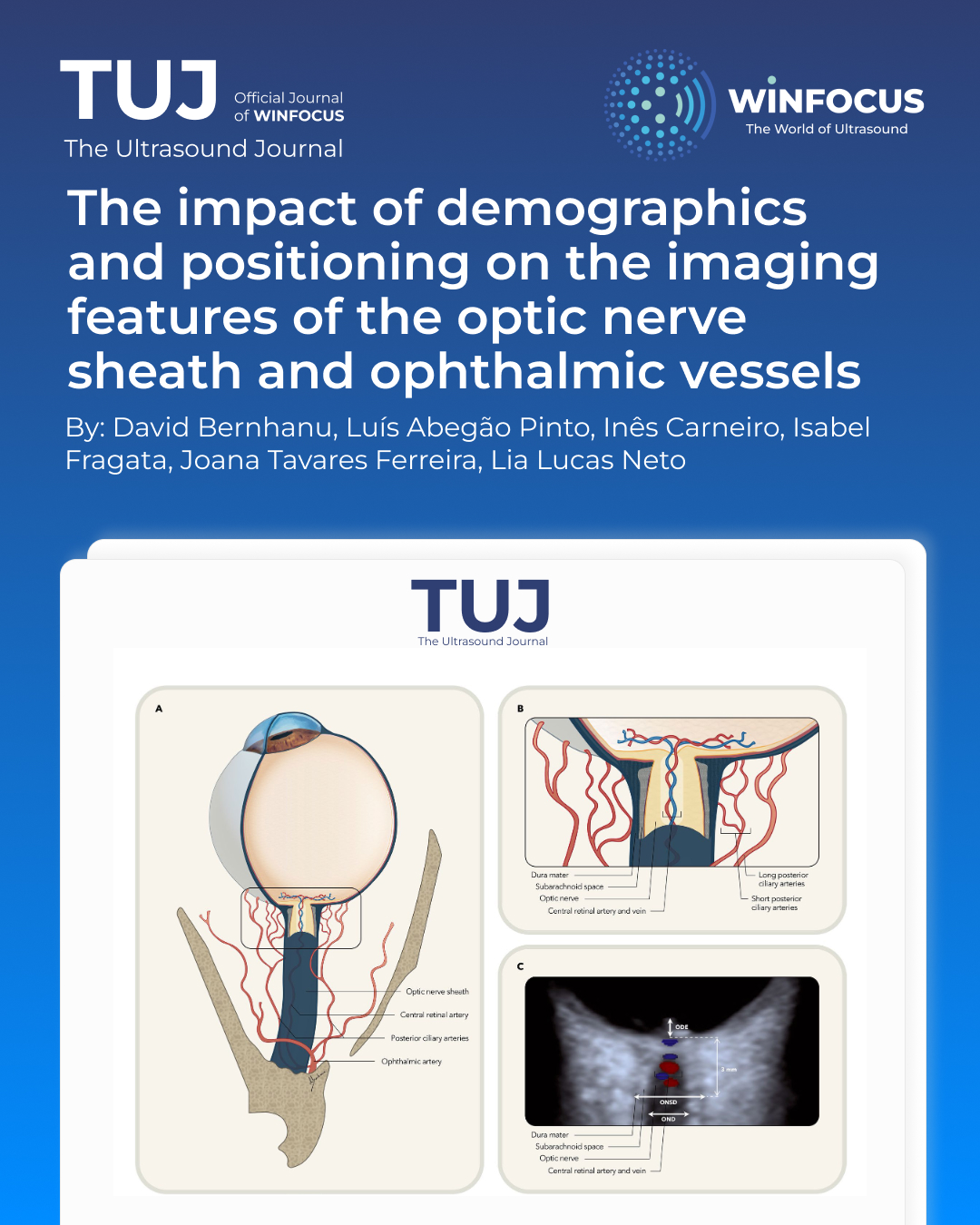
Optic nerve sheath, Ultrasonography, Doppler, Ophthalmic artery, Central retinal artery, Optic nerve sheath diameterAbstract
Background: There are significant discrepancies in the optic nerve sheath diameter (ONSD) reported in the literature. We aimed to determine the ultrasonographic imaging features of ONSD and ophthalmic vessels in a healthy population, using a standardized protocol, and to estimate the effect of demographics and positioning changes on imaging measurements.
Methods: We measured the mean values of the ONSD in supine and sitting position and the Doppler imaging parameters of the ophthalmic, central retinal and short posterior ciliary arteries. Inter-observer reliability was assessed using intraclass correlation coefficient (ICC). Linear regression models were fitted to predict the effect of demographic and clinical determinants on the imaging features.
Results: A total of 50 measurements were obtained for each observer. The mean ONSD was 5.9 mm and there was a mean reduction of 0.2 mm when assessed in sitting position (p < 0.001). Doppler analysis showed higher peak-systolic velocity and resistive index in the ophthalmic artery (35.6 cm/s vs. 12.0 cm/s; 0.78 vs. 0.70) compared to the central retinal artery (p < 0.001). Age, sex, heart rate and systolic blood pressure were significant determinants of the imaging features, with ONSD being larger in males (p < 0.001) and increasing with heart rate (p = 0.001). ICC estimates indicated ‘good’ inter-observer reliability of the ONSD and the ophthalmic and central retinal arteries velocities and resistance.
Conclusions: Our findings suggest a significant impact of patient demographics and positioning during ultrasonography on the normal imaging features of the ONSD and ophthalmic vessels. The heterogeneity in methodology and clinical cohorts may justify previous discrepancies in the literature. These findings can assist in the interpretation of imaging features in clinical settings and in the standardization of point of care ONSD ultrasonography.
References
1. Williamson TH, Harris A (1996) Color Doppler ultrasound imaging of the eye and orbit. Surv Ophthalmol 40:255–267
2. del Saz-Saucedo P, Redondo-Gonzalez O, Mateu-Mateu A, Huertas-Arroyo R, Garcia-Ruiz R, Botia-Paniagua E (2016) Sonographic assessment of the optic nerve sheath diameter in the diagnosis of idiopathic intracranial hypertension. J Neurol Sci 361:122–127
3. Chen Q, Chen W, Wang M, Sun X, Sha Y, Li Z, Tian G (2017) High-resolution transbulbar ultrasonography helping differentiate intracranial hypertension in bilateral optic disc oedema patients. Acta Ophthalmol 95:e481–e485
4. Ertl M, Barinka F, Torka E, Altmann M, Pfister K, Helbig H, Bogdahn U, Gamulescu MA, Schlachetzki F (2014) Ocular color-coded sonography—a promising tool for neurologists and intensive care physicians. Ultraschall Med 35:422–431
5. Koziarz A, Sne N, Kegel F, Nath S, Badhiwala JH, Nassiri F, Mansouri A, Yang K, Zhou Q, Rice T, Faidi S, Passos E, Healey A, Banfield L, Mensour M, Kirkpatrick AW, Nassar A, Fehlings MG, Hawryluk GWJ, Almenawer SA (2019) Bedside optic nerve ultrasonography for diagnosing increased intracranial pressure: a systematic review and meta-analysis. Ann Intern Med 171:896–905
6. Berhanu D, Ferreira JC, Abegao Pinto L, Aguiar de Sousa D, Lucas Neto L, Tavares Ferreira J (2023) The role of optic nerve sheath ultrasonography in increased intracranial pressure: a systematic review and meta analysis. J Neurol Sci 454:120853
7. Aletreby W, Alharthy A, Brindley PG, Kutsogiannis DJ, Faqihi F, Alzayer W, Balhahmar A, Soliman I, Hamido H, Alqahtani SA, Karakitsos D, Blaivas M (2022) Optic nerve sheath diameter ultrasound for raised intracranial pressure: a literature review and meta-analysis of its diagnostic accuracy. J Ultrasound Med 41:585–595
8. Dubourg J, Javouhey E, Geeraerts T, Messerer M, Kassai B (2011) Ultrasonography of optic nerve sheath diameter for detection of raised intracranial pressure: a systematic review and meta-analysis. Intensive Care Med 37:1059–1068
9. Wang L, Feng L, Yao Y, Wang Y, Chen Y, Feng J, Xing Y (2015) Optimal optic nerve sheath diameter threshold for the identification of elevated opening pressure on lumbar puncture in a Chinese population. PLoS ONE 10:e0117939
10. Agrawal D, Raghavendran K, Zhao L, Rajajee V (2020) A prospective study of optic nerve ultrasound for the detection of elevated intracranial pressure in severe traumatic brain injury. Crit Care Med 48:e1278–e1285 11. Sallam A, Abdelaal Ahmed Mahmoud MAA, Kamel MG, Hamza MK, Yassin HM, Hosny H, Younis MI, Ramadan E, Algameel HZ, Abdelhaq M, Abdelkader M, Mills KE, Mohamed H (2021) The diagnostic accuracy of noninvasive methods to measure the intracranial pressure: a systematic review and meta-analysis. Anesth Analg 132:686–695
12. Bauerle J, Schuchardt F, Schroeder L, Egger K, Weigel M, Harloff A (2013) Reproducibility and accuracy of optic nerve sheath diameter assessment using ultrasound compared to magnetic resonance imaging. BMC Neurol 13:187
13. Stevens RRF, Gommer ED, Aries MJH, Ertl M, Mess WH, Huberts W, Delhaas T (2021) Optic nerve sheath diameter assessment by neurosonology: a review of methodologic discrepancies. J Neuroimaging 31:814–825
14. Pansell J, Bell M, Rudberg P, Friman O, Cooray C (2022) Optic nerve sheath diameter measurement by ultrasound: evaluation of a standardized protocol. J Neuroimaging 32:104–110
15. Urbonas M, Raskauskiene N, Deltuva V, Bunevicius A (2022) Quantitative Evans index estimation using ultrasonographic measurement of the optic nerve sheath diameter in supine and upright position. Acta Neurochir 164:1755–1764
16. Xu X, Lu Y, Liu J, Xu R, Zhao K, Tao A (2023) Diagnostic value of the combination of ultrasonographic optic nerve sheath diameter and width of crural cistern with respect to the intracranial pressure in patients treated with decompressive craniotomy. Neurocrit Care 39:436–444
17. Berhanu D, Carneiro I, Antunes AP, Abegao Pinto L, Fragata I, Tavares Ferreira J, Lucas NL (2024) Dimensions of arachnoid bulk ratio: a superior optic nerve sheath index for intracranial pressure. Radiology 312:e240114 18. Ragauskas A, Matijosaitis V, Zakelis R, Petrikonis K, Rastenyte D, Piper I, Daubaris G (2012) Clinical assessment of noninvasive intracranial pressure absolute value measurement method. Neurology 78:1684–1691
19. Tarzamni MK, Derakhshan B, Meshkini A, Merat H, Fouladi DF, Mostafazadeh S, Rezakhah A (2016) The diagnostic performance of ultrasonographic optic nerve sheath diameter and color Doppler indices of the ophthalmic arteries in detecting elevated intracranial pressure. Clin Neurol Neurosurg 141:82–88
20. Jeub M, Schlapakow E, Ratz M, Kindler C, Schievelkamp AH, Wabbels B, Kornblum C (2020) Sonographic assessment of the optic nerve and the central retinal artery in idiopathic intracranial hypertension. J Clin Neurosci 72:292–297
21. Querfurth HW, Lagreze WD, Hedges TR, Heggerick PA (2002) Flow velocity and pulsatility of the ocular circulation in chronic intracranial hypertension. Acta Neurol Scand 105:431–440
22. Abegao Pinto L, Vandewalle E, De Clerck E, Marques-Neves C, Stalmans I (2012) Ophthalmic artery Doppler waveform changes associated with increased damage in glaucoma patients. Invest Ophthalmol Vis Sci 53:2448–2453
23. Galassi F, Nuzzaci G, Sodi A, Casi P, Vielmo A (1992) Color Doppler imaging in evaluation of optic nerve blood supply in normal and glaucomatous subjects. Int Ophthalmol 16:273–276
24. Tranquart F, Berges O, Koskas P, Arsene S, Rossazza C, Pisella PJ, Pourcelot L (2003) Color Doppler imaging of orbital vessels: personal experience and literature review. J Clin Ultrasound 31:258–273
25. Koo TK, Li MY (2016) A guideline of selecting and reporting intraclass correlation coefficients for reliability research. J Chiropr Med 15:155–163
26. Gisev N, Bell JS, Chen TF (2013) Interrater agreement and interrater reliability: key concepts, approaches, and applications. Res Social Adm Pharm 9:330–338
27. Rajajee V, Vanaman M, Fletcher JJ, Jacobs TL (2011) Optic nerve ultrasound for the detection of raised intracranial pressure. Neurocrit Care 15:506–515
28. Schroeder C, Katsanos AH, Richter D, Tsivgoulis G, Gold R, Krogias C (2020) Quantification of optic nerve and sheath diameter by transorbital sonography: a systematic review and metanalysis. J Neuroimaging 30:165–174
29. Weigel M, Lagreze WA, Lazzaro A, Hennig J, Bley TA (2006) Fast and quantitative high-resolution magnetic resonance imaging of the optic nerve at 3.0 tesla. Invest Radiol 41:83–86
30. Abegao Pinto L, Vandewalle E, Pronk A, Stalmans I (2012) Intraocular pressure correlates with optic nerve sheath diameter in patients with normal tension glaucoma. Graefes Arch Clin Exp Ophthalmol 250:1075–1080
31. Jaggi GP, Miller NR, Flammer J, Weinreb RN, Remonda L, Killer HE (2012) Optic nerve sheath diameter in normal-tension glaucoma patients. Br J Ophthalmol 96:53–56
32. Berhanu D, Carneiro I, Abegão Pinto L, Tavares Ferreira J, Lucas NL (2022) The optic nerve-sheath anatomy in intracranial pressure [abstract]. In: 13° Simposio Internacional de Anatomía Clínica y Aplicada. Rev Argent Anat Clin 14(3):123–144
33. Nabeta HW, Bahr NC, Rhein J, Fossland N, Kiragga AN, Meya DB, Dunlop SJ, Boulware DR (2014) Accuracy of noninvasive intraocular pressure or optic nerve sheath diameter measurements for predicting elevated intracranial pressure in cryptococcal meningitis. Open Forum Infect Dis 1:ofu093
34. Mohson KI, Auday N (2019) Role of orbital ultrasound in the assessment of clinically detected papilledema. J Med Ultrasound 27:135–140
35. Blaivas M, Theodoro D, Sierzenski PR (2003) Elevated intracranial pressure detected by bedside emergency ultrasonography of the optic nerve sheath. Acad Emerg Med 10:376–381
36. Tayal VS, Neulander M, Norton HJ, Foster T, Saunders T, Blaivas M (2007) Emergency department sonographic measurement of optic nerve sheath diameter to detect findings of increased intracranial pressure in adult head injury patients. Ann Emerg Med 49:508–514
37. Goel RS, Goyal NK, Dharap SB, Kumar M, Gore MA (2008) Utility of optic nerve ultrasonography in head injury. Injury 39:519–524
38. Major R, Girling S, Boyle A (2011) Ultrasound measurement of optic nerve sheath diameter in patients with a clinical suspicion of raised intracranial pressure. Emerg Med J 28:679–681
39. Qayyum H, Ramlakhan S (2013) Can ocular ultrasound predict intracranial hypertension? A pilot diagnostic accuracy evaluation in a UK emergency department. Eur J Emerg Med 20:91–97
40. Golshani K, Ebrahim Zadeh M, Farajzadegan Z, Khorvash F (2015) Diagnostic accuracy of optic nerve ultrasonography and ophthalmoscopy in prediction of elevated intracranial pressure. Emergency 3:54–58
41. Kaur A, Gautam PL, Sharma S, Singh VP, Sharma S (2021) Bedside ultrasonographic assessment of optic nerve sheath diameter as a means of detecting raised intracranial pressure in neuro-trauma patients: a crosssectional study. Ann Indian Acad Neurol 24:63–68
42. Pansell J, Bell M, Rudberg P, Friman O, Cooray C (2023) Optic nerve sheath diameter in intracranial hypertension: measurement external or internal of the dura mater? J Neuroimaging 33:58–66
43. Ertl M, Knuppel C, Veitweber M, Wagner A, Pfister K, Wendl C, Baldaranov D, Beck J, Linker RA, Schlachetzki F (2020) Normal age- and sex-related values of the optic nerve sheath diameter and its dependency on position and positive end-expiratory pressure. Ultrasound Med Biol 46:3279–3285
44. Bauerle J, Lochner P, Kaps M, Nedelmann M (2012) Intra- and interobsever reliability of sonographic assessment of the optic nerve sheath diameter in healthy adults. J Neuroimaging 22:42–45
45. Oberfoell S, Murphy D, French A, Trent S, Richards D (2017) Inter-rater reliability of sonographic optic nerve sheath diameter measurements by emergency medicine physicians. J Ultrasound Med 36:1579–1584
46. Cardim D, Czosnyka M, Chandrapatham K, Badenes R, Bertuccio A, Noto AD, Donnelly J, Pelosi P, Ball L, Hutchinson PJ, Robba C (2020) Effects of age and sex on optic nerve sheath diameter in healthy volunteers and patients with traumatic brain injury. Front Neurol 11:764
47. Goeres P, Zeiler FA, Unger B, Karakitsos D, Gillman LM (2016) Ultrasound assessment of optic nerve sheath diameter in healthy volunteers. J Crit Care 31:168–171
48. Pansell J, Rudberg PC, Friman O, Bell M, Cooray C (2024) Sex differences in the diagnostic value of optic nerve sheath diameter for assessing intracranial pressure. Sci Rep 14:9553
49. Dimitri GM, Agrawal S, Young A, Donnelly J, Liu X, Smielewski P, Hutchinson P, Czosnyka M, Lio P, Haubrich C (2018) Simultaneous transients of intracranial pressure and heart rate in traumatic brain injury: methods of analysis. Acta Neurochir Suppl 126:147–151
50. Luis A, Santos AS, Dias C, Almeida R, Rocha AP (2015) Heart rate variability during plateau waves of intracranial pressure: a pilot descriptive study. Annu Int Conf IEEE Eng Med Biol Soc 2015:6142–6145
51. Cui W, Roberson DA, Chen Z, Madronero LF, Cuneo BF (2008) Systolic and diastolic time intervals measured from Doppler tissue imaging: normal values and Z-score tables, and effects of age, heart rate, and body surface area. J Am Soc Echocardiogr 21:361–370
52. Sarnari R, Kamal RY, Friedberg MK, Silverman NH (2009) Doppler assessment of the ratio of the systolic to diastolic duration in normal children: relation to heart rate, age and body surface area. J Am Soc Echocardiogr 22:928–932
53. Bradley WG Jr (2015) CSF flow in the brain in the context of normal pressure hydrocephalus. AJNR Am J Neuroradiol 36:831–838
54. Bradley WG Jr, Kortman KE, Burgoyne B (1986) Flowing cerebrospinal fluid in normal and hydrocephalic states: appearance on MR images. Radiology 159:611–616
55. Kaiser HJ, Schotzau A, Flammer J (1996) Blood-flow velocities in the extraocular vessels in normal volunteers. Am J Ophthalmol 122:364–370
56. Guthoff RF, Berger RW, Winkler P, Helmke K, Chumbley LC (1991) Doppler ultrasonography of the ophthalmic and central retinal vessels. Arch Ophthalmol 109:532–536
57. Karami M, Shirazinejad S, Shaygannejad V, Shirazinejad Z (2015) Transocular Doppler and optic nerve sheath diameter monitoring to detect intracranial hypertension. Adv Biomed Res 4:231
58. Wiacek MP, Modrzejewska M, Zaborski D (2020) Age-related changes in retrobulbar circulation: a literature review. Int Ophthalmol 40:493–501 59. Gillies WE, Brooks AM, Scott M, Ryan L (1999) Comparison of colour Doppler imaging of orbital vessels in elderly compared with young adult patients. Aust N Z J Ophthalmol 27:173–175
60. Dellafiore C, Lava M, Verticchio Vercellin AC, Cutolo CA, Tinelli C, De Silvestri A, Calliada F, Milano G (2015) Reproducibility of retrobulbar blood flow velocity measurements in normal subjects using two different CDI devices. Radiol Med 120:737–744
61. Vercellin Alice CV, Cutolo CA, Dellafiore C, Lava M, Tinelli C, De Silvestri A, Calliada F, Milano G (2016) Inter-device reproducibility of retrobulbar blood flow velocity measurements in healthy subjects using color Doppler imaging. J Ultrasound 19:125–130
Downloads
Published
Issue
Section
License
Copyright (c) 2025 David Bernhanu, Luís Abegão Pinto, Inês Carneiro, Isabel Fragata, Joana Tavares Ferreira, Lia Lucas Neto (Author)

This work is licensed under a Creative Commons Attribution-NonCommercial 4.0 International License.
This is an Open Access article distributed under the terms of the Creative Commons Attribution License (https://creativecommons.org/licenses/by-nc/4.0) which permits unrestricted use, distribution, and reproduction in any medium, provided the original work is properly cited.
Authors retain the copyright for their published work. No formal permission will be required to reproduce parts (tables or illustrations) of published papers, provided the source is quoted appropriately and reproduction has no commercial intent.
How to Cite


