
Prediction of post-extubation stridor using ultrasound measurement of subglottic airway diameter
Keywords:
point-of-care ultrasound, critical care, Pediatrics, post extubation stridorAbstract
Background: Post-extubation stridor (PES) is a common complication in children in the pediatric intensive care unit (PICU). Despite the evaluation of various predictive tests, no single method has consistently demonstrated strong predictive value for PES. We evaluated the diagnostic utility of subglottic diameter ultrasonography for predicting PES within 48-h post-extubation in children.
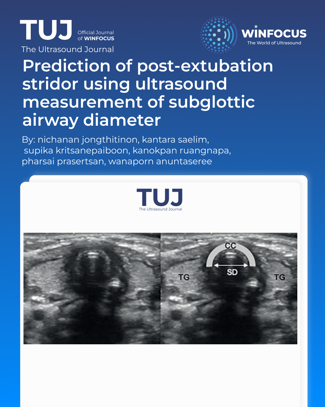
Material and methods: The prospective observational study in university-affiliated PICU was done in children aged 1 month to 15 years who successfully passed a spontaneous breathing trial (SBT) and were scheduled for extubation were enrolled between January 2024 and January 2025. During SBT and cuff deflation, subglottic ultrasonography was performed at a peak inspiratory pressure (PIP) of 15 cmH2O, and the subglottic ratio (SR) and intracricoid peritubular free space (IPFS) were calculated. Leak percentage was calculated as the ratio of the difference between inspiratory and expiratory volumes over inspiratory volume at each PIP level.
Results: PES occurred in 33 of 176 (18.75%) patients. In children aged ≥1 year, SR was significantly associated with PES (adjusted OR, 3.78; 95% CI: 1.28–12.21). An SR <1.30 showed 70% sensitivity, 65% specificity, and
66% accuracy. IPFS <2.08 mm demonstrated 80% sensitivity, 65% specificity, and 68% accuracy. A leak percentage <36.77% at 25 cmH2O yielded 80% sensitivity, 47% specificity, and 53% accuracy. AUC values for SR, IPFS, and leak percentage were 0.664, 0.691, and 0.648, respectively.
Conclusion: Among the evaluated parameters, IPFS showed the highest diagnostic performance for predicting PES, slightly outperforming the subglottic ratio in patients aged ≥1 year.
References
1. El Amrousy D, Elkashlan M, Elshmaa N, Ragab A. -Ultrasound-Guided Laryngeal Air Column Width Difference as a New Predictor for Postextubation Stridor in Children. Crit Care Med. 2018 Jun;46(6):e496-e501. doi: 10.1097/CCM.0000000000003068
2. Samprathi M, Baranwal AK, Gupta PK, Jayashree M. Pre-extubation ultrasonographic measurement of intracricoid peritubal free space: A pilot study to predict post-extubation airway obstruction in children. Int J Pediatr Otorhinolaryngol. 2020 Nov;138:110348. doi: 10.1016/j.ijporl.2020.110348
3. Kanno K, Fujiwara N, Moromizato T, Fujii S, Ami Y, Tokushige A, et al. S. Pre-Extubation Cuffed Tube Leak Test and Subsequent Post-Extubation Laryngeal Edema: -Prospective, Single-Center Evaluation of PICU Patients. Pediatr Crit Care Med. 2023 Sep 1;24(9):767-74. doi: 10.1097/PCC.0000000000003282
4. Girard TD, Alhazzani W, Kress JP, Ouellette DR, Schmidt GA, Truwit JD, et al. An Official American Thoracic Society/American College of Chest Physicians Clinical Practice Guideline: Liberation from Mechanical Ventilation in Critically Ill Adults. Rehabilitation Protocols, Ventilator Liberation Protocols, and Cuff Leak Tests. Am J Respir Crit Care Med. 2017 Jan 1;195(1):120-33. doi: 10.1164/rccm.201610-2075ST
5. Ochoa ME, Marín Mdel C, Frutos-Vivar F, Gordo F, Latour-Pérez J, Calvo E, Esteban A. Cuff-leak test for the diagnosis of upper airway obstruction in adults: a systematic review and meta-analysis. Intensive Care Med. 2009 Jul;35(7):1171-9. doi: 10.1007/s00134-009-1501-9
6. Kuriyama A, Jackson JL, Kamei J. Performance of the cuff leak test in adults in predicting post-extubation airway complications: a systematic review and meta-analysis. Crit Care. 2020 Nov 7;24(1):640. doi: 10.1186/s13054-020-03358-8
7. Bhargava T, Kumar A, Bharti A, Khuba S. Comparison of Laryngeal Ultrasound and Cuff Leak Test to Predict Post-Extubation Stridor in Total Thyroidectomy. Turk J Anaesthesiol Reanim. 2021 Jun;49(3):238-43. doi: 10.5152/TJAR.2020.176
8. Strauss S. Sonographic appearance of cricoid cartilage calcification in healthy children. AJR Am J Roentgenol. 2000 Jan;174(1):223-8. doi: 10.2214/ajr.174.1.1740223
9. Johnson DW. Croup. BMJ Clin Evid. 2014 Sep 29;2014:0321. PMID: 25263284
10. Kimura S, Ahn JB, Takahashi M, Kwon S, Papatheodorou S. Effectiveness of corticosteroids for post-extubation stridor and extubation failure in pediatric patients: a systematic review and meta-analysis. Ann Intensive Care. 2020 Nov 18;10(1):155. doi: 10.1186/s13613-020-00773-6
11. Burton L, Loberger J, Baker M, Prabhakaran P, Bhargava V. Pre-Extubation Ultrasound Measurement of In Situ Cuffed Endotracheal Tube Laryngeal Air Column Width Difference: Single-Center Pilot Study of Relationship With Post-Extubation Stridor in Subjects Younger Than 5 Years Old. Pediatr Crit Care Med. 2024 Mar 1;25(3):222-30. doi: 10.1097/PCC.0000000000003377
12. Vijayasekaran S. Pediatric Airway Pathology. Front Pediatr. 2020 Jun 4;8:246. doi: 10.3389/fped.2020.00246
13. Topjian AA, Raymond TT, Atkins D, Chan M, Duff JP, Joyner BL, et al. Part 4: Pediatric Basic and Advanced Life Support 2020 American Heart Association Guidelines for Cardiopulmonary Resuscitation and Emergency Cardiovascular Care. Pediatrics. 2021 Jan;147(Suppl 1):e2020038505D. doi: 10.1542/peds.2020-038505D
14. Emeriaud G, Harrington K, Jouvet P. Diagnosis of Post--extubation Stridor: Easier with Technology Support? Am J Respir Crit Care Med. 2016 Jan 15;193(2):113-5. doi: 10.1164/rccm.201509-1905ED
15. Konca C, Tekin M, Kucuk A. Incidence of Mechanical Ventilation Adverse Events in Critically Ill Children in a Tertiary Pediatric Intensive Care Unit. Turk Thorac J. 2022 Jul;23(4):277-83. doi: 10.5152/TurkThoracJ.2022.21253
16. Principi T, Fraser DD, Morrison GC, Farsi SA, Carrelas JF, Maurice EA, et al. Complications of mechanical ventilation in the pediatric population. Pediatr Pulmonol. 2011 May;46(5):452-7. doi: 10.1002/ppul.21389
17. Chand R, Roy Chowdhury S, Rupert E, Mandal CK, Narayan P. Benefits of Using High-Volume-Low-Pressure Tracheal Tube in Children Undergoing Congenital Cardiac Surgery: Evidence From a Prospective Randomized Study. Semin Cardiothorac Vasc Anesth. 2018 Sep;22(3):300-5. doi: 10.1177/1089253217750753
Downloads
Published
Issue
Section
License
Copyright (c) 2026 Nichanan Jongthitinon, Kantara Saelim, Supika Kritsanepaiboon, Kanokpan Ruangnapa, Pharsai Prasertsan, Wanaporn Anuntaseree (Author)

This work is licensed under a Creative Commons Attribution-NonCommercial 4.0 International License.
This is an Open Access article distributed under the terms of the Creative Commons Attribution License (https://creativecommons.org/licenses/by-nc/4.0) which permits unrestricted use, distribution, and reproduction in any medium, provided the original work is properly cited.
Authors retain the copyright for their published work. No formal permission will be required to reproduce parts (tables or illustrations) of published papers, provided the source is quoted appropriately and reproduction has no commercial intent.
How to Cite


