
Lung ultrasound for etiological diagnosis of pneumonia in the emergency department: correlation with bronchoalveolar lavage results
Keywords:
Lung ultrasound, Molecular syndromic panels, PCR, Bronchoalveolar lavage, BronchoscopyAbstract
Background: Pneumonia is the leading cause of death from infectious diseases worldwide. Lung ultrasound (LUS) is highly accurate for chest infections diagnosis, yet its correlation with causative pathogens remains unclear. Respiratory cultures, combined with molecular techniques represent the gold standard, achieving etiological diagnosis in 90–95% of cases. We compared LUS findings with bronchoalveolar lavage (BAL) sample analyses; to our knowledge, no prior studies have investigated this in the emergency department (ED).
Materials and methods: Bronchoalveolar lavage (BAL)-LUS is a prospective observational non-profit study conducted in the ED, aiming to assess whether there is a correlation between the LUS sonographic appearance, assessed blindly across 12 lung fields, and the etiopathogenetic agent of pneumonia (bacterial and viral) detected with molecular syndromic panels (MSPs) and respiratory cultures obtained with BAL.
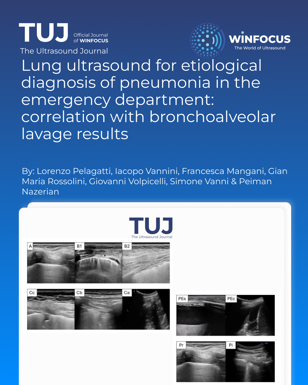
Results: 64 patients were enrolled (mean age 73.3 ± 14.6) with 11 diagnosed as viral pneumonia and 53 as bacterial pneumonia. Bacterial pneumonias were more commonly associated with consolidation (2.9 ± 2.2 vs. 1.5 ± 0.9, p < 0.01) and a higher incidence of pleural effusion (0.9 ± 1.3 vs. 0.3 ± 0.6, p < 0.01). Viral pneumonias were more often associated with interstitial syndrome (4.9 ± 3.3 vs. 0.5 ± 1.3, p < 0.01) and small subpleural consolidations (0.9 ± 1.8 vs. 0.2 ± 0.6, p = 0.01). The mean LUS score was significantly higher in bacterial than in viral pneumonia with a AUC of 0.81 (95% CI 0.68–0.93).
Conclusions: Viral pneumonia is usually associated with interstitial syndrome and small subpleural consolidations; on the other hand, bacterial pneumonia is usually associated with consolidation, and pleural effusion.
References
1. Torres A, Niederman MS, Chastre J, Ewig S, Fernandez-Vandellos P, Hanberger H, Kollef M, Li Bassi G, Luna CM, Martin-Loeches I, Paiva JA, Read RC, Rigau D, Timsit JF, Welte T, Wunderink R, International (2017) ERS/ESICM/ESCMID/ALAT guidelines for the management of hospital-acquired pneumonia and ventilator-associated pneumonia: guidelines for the management of hospital-acquired pneumonia (HAP)/ventilator-associated pneumonia (VAP) of the European respiratory society (ERS), European society of intensive care medicine (ESICM), European society of clinical microbiology and infectious diseases (ESCMID) and Asociación Latinoamericana Del Tórax (ALAT). Eur Respir J 50(3):1700582. https://doi.org/10.1183/13993003.00582-2017
2. Ramirez JA, Wiemken TL, Peyrani P, Arnold FW, Kelley R, Mattingly WA, Nakamatsu R, Pena S, Guinn BE, Furmanek SP, Persaud AK, Raghuram A, Fernandez F, Beavin L, Bosson R, Fernandez-Botran R, Cavallazzi R, Bordon J, Valdivieso C, Schulte J, Carrico RM (2017) University of Louisville pneumonia study Group. Adults hospitalized with pneumonia in the united states: Incidence, Epidemiology, and mortality. Clin Infect Dis 65(11):1806–1812. https://doi.org/10.1093/cid/cix647
3. American Thoracic Society; Infectious Diseases Society of America (2005) Guidelines for the management of adults with hospital-acquired, ventilator-associated, and healthcare-associated pneumonia. Am J Respir Crit Care Med 171(4):388–416. https://doi.org/10.1164/rccm.200405-644ST
4. Nazerian P, Volpicelli G, Vanni S, Gigli C, Betti L, Bartolucci M, Zanobetti M, Ermini FR, Iannello C, Grifoni S (2015) Accuracy of lung ultrasound for the diagnosis of consolidations when compared to chest computed tomography. Am J Emerg Med 33(5):620–625. https://doi.org/10.1016/j.ajem.2015.01.035
5. Haaksma ME, Smit JM, Heldeweg MLA, Nooitgedacht JS, de Grooth HJ, Jonkman AH, Girbes ARJ, Heunks L, Tuinman PR (2022) Extended lung ultrasound to differentiate between pneumonia and atelectasis in critically ill patients: A diagnostic accuracy study. Crit Care Med 50(5):750–759. https://doi.org/10.1097/CCM.0000000000005303
6. Ye X, Xiao H, Chen B, Zhang S (2015) Accuracy of lung ultrasonography versus chest radiography for the diagnosis of adult Community-Acquired pneumonia: review of the literature and Meta-Analysis. PLoS ONE 10(6):e0130066. https://doi.org/10.1371/journal.pone.0130066
7. Laursen CB, Sloth E, Lassen AT, Christensen Rd, Lambrechtsen J, Madsen PH, Henriksen DP, Davidsen JR, Rasmussen F (2014) Point-of-care ultrasonography in patients admitted with respiratory symptoms: a single-blind, randomised controlled trial. Lancet Respir Med 2(8):638–646. https://doi.org/10.1016/S2213-2600(14)70135-3
8. Escudero D, Fernández-Suarez J, Forcelledo L, Balboa S, Fernández J, Astola I, Quindos B, Campos R, Vázquez F, Boga JA (2022) Evaluation and clinical impact of biofire filmarray pneumonia panel plus in ICU-hospitalized COVID-19 patients. Diagnostics (Basel) 12(12):3134. https://doi.org/10.3390/diagnostics12123134
9. Volpicelli G, Elbarbary M, Blaivas M, Lichtenstein DA, Mathis G, Kirkpatrick AW, Melniker L, Gargani L, Noble VE, Via G, Dean A, Tsung JW, Soldati G, Copetti R, Bouhemad B, Reissig A, Agricola E, Rouby JJ, Arbelot C, Liteplo A, Sargsyan A, Silva F, Hoppmann R, Breitkreutz R, Seibel A, Neri L, Storti E, Petrovic T (2012) International Liaison Committee on Lung Ultrasound (ILC-LUS) for International Consensus Conference on Lung Ultrasound (ICC-LUS). International evidence-based recommendations for point-of-care lung ultrasound. Intensive Care Med 38(4):577–91. https://doi.org/10.1007/s00134-012-2513-4
10. Bouhemad B, Liu ZH, Arbelot C, Zhang M, Ferarri F, Le-Guen M, Girard M, Lu Q, Rouby JJ (2010) Ultrasound assessment of antibiotic-induced pulmonary reaeration in ventilator-associated pneumonia. Crit Care Med 38(1):84–92. https://doi.org/10.1097/CCM.0b013e3181b08cdb
11. Klech H, Pohl W (1989) Technical recommendations and guidelines for Bronchoalveolar lavage (BAL). Eur Respir J 2:561–585
12. Zhu F, Zhao X, Wang T et al (2021) Ultrasonic characteristics and severity assessment of lung ultrasound in COVID-19 pneumonia in Wuhan, china: A Retrospective, observational study. Eng (Beijing) 7(3):367–375. https://doi.org/10.1016/j.eng.2020.09.007
13. Haaksma ME, Smit JM, Heldeweg MLA et al (2022) Extended lung ultrasound to differentiate between pneumonia and atelectasis in critically ill patients: A diagnostic accuracy study. Crit Care Med 50(5):750–759. https://doi.org/10.1097/CCM.0000000000005303
14. Guitart C, Rodríguez-Fanjul J, Bobillo-Perez S et al (2022) An algorithm combining procalcitonin and lung ultrasound improves the diagnosis of bacterial pneumonia in critically ill children: the PROLUSP study, a randomized clinical trial. Pediatr Pulmonol 57(3):711–723. https://doi.org/10.1002/ppul.25790
15. Malla D, Rathi V, Gomber S, Upreti L (2021) Can lung ultrasound differentiate between bacterial and viral pneumonia in children? J Clin Ultrasound 49(2):91–100. https://doi.org/10.1002/jcu.22951
16. Stoicescu ER et al (2024) Differentiating viral from bacterial pneumonia in children: the diagnostic role of lung Ultrasound—A prospective observational study. Diagnostics 14:480
17. Boccatonda A, Cocco G, D’Ardes D et al (2023) Infectious pneumonia and lung ultrasound: A review. J Clin Med 12(4):1402. https://doi.org/10.3390/jcm12041402
18. Beshara M, Bittner EA, Goffi A, Berra L, Chang MG (2024) Nuts and bolts of lung ultrasound: utility, scanning techniques, protocols, and findings in common pathologies. Crit Care 28(1):328. https://doi.org/10.1186/s13054-024-05102-y
19. Vollmer I (2021) Thoracic ultrasound in viral infections. Ecografía torácica de Las infecciones víricas. Radiologia (Engl Ed) 63(3):252–257. https://doi.org/10.1016/j.rx.2020.12.005
20. Bitar ZI, Shamsah M, Maadarani O, Bamasood OM, Bitar AZ, Alfoudri H (2021) Lung ultrasound and sonographic subpleural consolidation in COVID-19 pneumonia correlate with disease severity. Crit Care Res Pract 2021:6695033. https://doi.org/10.1155/2021/6695033
21. Volpicelli G, Gargani L, Perlini S, Spinelli S, Barbieri G, Lanotte A, Casasola GG, Nogué-Bou R, Lamorte A, Agricola E, Villén T, Deol PS, Nazerian P, Corradi F, Stefanone V, Fraga DN, Navalesi P, Ferre R, Boero E, Martinelli G, Cristoni L, Perani C, Vetrugno L, McDermott C, Miralles-Aguiar F, Secco G, Zattera C, Salinaro F, Grignaschi A, Boccatonda A, Giostra F, Infante MN, Covella M, Ingallina G, Burkert J, Frumento P, Forfori F, Ghiadoni L, on behalf of the International Multicenter Study Group on LUS (2021) COVID-19. Lung ultrasound for the early diagnosis of COVID-19 pneumonia: an international multicenter study. Intensive Care Med 47(4):444–454. https://doi.org/10.1007/s00134-021-06373-7
22. Millington SJ, Koenig S, Mayo P, Volpicelli G (2021) Lung ultrasound for patients with coronavirus disease 2019 pulmonary disease. Chest 159(1):205–211. https://doi.org/10.1016/j.chest.2020.08.2054
23. Volpicelli G, Lamorte A, Villén T (2020) What’s new in lung ultrasound during the COVID-19 pandemic. Intensive Care Med 46(7):1445–1448. https://doi.org/10.1007/s00134-020-06048-9
24. Tung-Chen Y, Giraldo Hernández A, Mora Vargas A et al (2022) Impact of lung ultrasound during the SARS-CoV-2 pandemic: distinction between viral and bacterial pneumonia. Reumatol Clin (Engl Ed) 18(9):546–550. https://doi.org/10.1016/j.reumae.2021.09.006
25. Bianchi S, Savinelli C, Paolucci E et al (2022) Point-of-care ultrasound (PoCUS) in the early diagnosis of novel coronavirus 2019 disease (COVID-19) in a first-level emergency department during a SARS-CoV-2 outbreak in italy: a real-life analysis. Intern Emerg Med 17(1):193–204. https://doi.org/10.1007/s11739-021-02643-w
26. Barbieri G, De Vuono S, Gargani L, Berisha S, Spinelli S, Del Carlo C, Deri C, D’Angelo G, Groff P, Ghiadoni L (2024) Prognostic value of lung ultrasound score performed in the emergency department in COVID-19 patients: A prospective multicenter study in central Italy. Emerg Care J 20(1):12268
27. Kamat IS, Ramachandran V, Eswaran H, Guffey D, Musher DM (2020) Procalcitonin to distinguish viral from bacterial pneumonia: A systematic review and Meta-analysis. Clin Infect Dis 70(3):538–542. https://doi.org/10.1093/cid/ciz545
28. Nazerian P, Cerini G, Vanni S et al (2016) Diagnostic accuracy of lung ultrasonography combined with procalcitonin for the diagnosis of pneumonia: a pilot study. Crit Ultrasound J 8(1):17. https://doi.org/10.1186/s13089-016-0054-8
29. Omran A, Awad H, Ibrahim M, El-Sharkawy S, Elfiky S, Rezk AR (2022) Lung ultrasound and neutrophil lymphocyte ratio in early diagnosis and differentiation between viral and bacterial pneumonia in young children. Child (Basel) 9(10):1457. https://doi.org/10.3390/children9101457
30. Jain S, Self WH, Wunderink RG et al (2015) Community-Acquired pneumonia requiring hospitalization among U.S. Adults. N Engl J Med 373(5):415–427. https://doi.org/10.1056/NEJMoa1500245
31. Niederman MS, Torres A (2022) Severe community-acquired pneumonia. Eur Respir Rev 31(166):220123 Published 2022 Dec 14. https://doi.org/10.1183/16000617.0123-2022
32. Cilloniz C, Ewig S, Gabarrus A et al (2017) Seasonality of pathogens causing community-acquired pneumonia. Respirology 22(4):778–785. https://doi.org/10.1111/resp.12978
33. Lim YK, Kweon OJ, Kim HR, Kim TH, Lee MK (2019) Impact of bacterial and viral coinfection in community-acquired pneumonia in adults. Diagn Microbiol Infect Dis 94(1):50–54. https://doi.org/10.1016/j.diagmicrobio.2018.11.014
34. Ruuskanen O, Lahti E, Jennings LC, Murdoch DR (2011) Viral pneumonia. Lancet 377(9773):1264–1275. https://doi.org/10.1016/S0140-6736(10)61459-6
Downloads
Published
Issue
Section
License
Copyright (c) 2025 Lorenzo Pelagatti, Iacopo Vannini, Francesca Mangani, Gian Maria Rossolini, Giovanni Volpicelli, Simone Vanni, Peiman Nazerian (Author)

This work is licensed under a Creative Commons Attribution-NonCommercial 4.0 International License.
This is an Open Access article distributed under the terms of the Creative Commons Attribution License (https://creativecommons.org/licenses/by-nc/4.0) which permits unrestricted use, distribution, and reproduction in any medium, provided the original work is properly cited.
Authors retain the copyright for their published work. No formal permission will be required to reproduce parts (tables or illustrations) of published papers, provided the source is quoted appropriately and reproduction has no commercial intent.
How to Cite


