
Ultrasound-based statistical shape modeling for prognosis in unstable hip dysplasia
Keywords:
Ultrasound, Neonatal, DDH, Unstable, Dysplasia, Hip, SSM, Statistical shape modelingAbstract
Background: Current methods to classify developmental dysplasia of the hip (DDH) on ultrasound (US) images, such as the Graf method, provide limited prognostic information. This study aimed to improve the prediction of the clinical course and outcome at age five of decentered hips, diagnosed on the first US made in the first months after birth, by identifying acetabular shape variants on these US images using a statistical shape model (SSM).
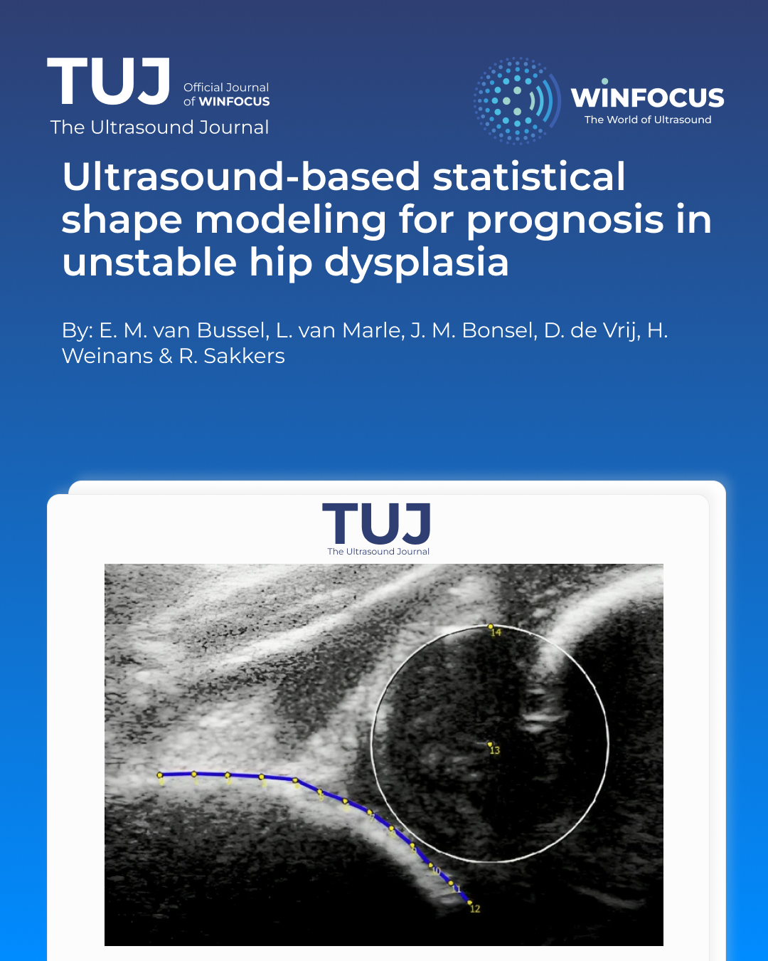
Patients and Methods: US images of the hip were retrieved from a single-center retrospective cohort of patients with DDH Graf type D/III/IV. A SSM was created from the US images made at initial diagnosis.. The association between the identified acetabular shape variants and an unfavorable outcome (residual DDH at age five and open reduction and/or a pelvic osteotomy before age five) was established with multivariable regression models.
Results: 92 decentered dysplastic hips with full history could be retrieved from the database and were included. At age five, 12 patients (13%) had undergone open reduction, 13 (14%) had a pelvic osteotomy, and 32 (35%) patients showed residual DDH. Four shape variants represented 95% of the variance in acetabular shape. Mode 4 was associated with an unfavorable outcome (odds ratio (OR): 1.80 (95% CI 1.12–2.90). Mode 1 was associated with less risk on open reductions or pelvic osteotomies (OR: 0.56 (95% CI 0.33–0.96).
Conclusions: A potential new method of analyzing US images for DDH using SSM established four distinct acetabular shapes on neonatal US images with unstable DDH, of which two were associated with outcomes at five years of age. This tool could serve as a basis for a better prediction of outcome and a more personalized and effective guide for treatment.
References
1. Ambellan F, Lamecker H, von Tycowicz C, Zachow S (2019) Statistical shape models: understanding and mastering variation in anatomy. Adv Exp Med Biol 1156:67–84. https:// doi. org/ 10. 1007/ 978-3- 030- 19385-0_5
2. Baumann T, Gather K, Pavone V et al (2022) Four decades of developmental dysplastic hip screening according to graf what have we learned. Front Pediatr. https:// doi. org/ 10. 3389/ fped. 2022. 990806
3. Bonsel JM, Gielis WP, Pollet V et al (2022) Statistical shape modeling of US images to predict hip dysplasia development in infants. Radiology 303:425–432. https:// doi. org/ 10. 1148/ RADIOL. 211057
4. Bozkurt C, Bekin Sarikaya PZ, Kaptan AY et al (2022) Predictivity of international hip dysplasia institute classification in pavlik harness treatment and correlation with graf ultrasonographic classification. J Pediatr Orthop B 31:232–236. https:// doi. org/ 10. 1097/ BPB. 00000 00000 000876
5. Cai X, Wu Y, Huang J et al (2024) Application of statistical shape models in orthopedics: a narrative review. Intell Med 4:249–255. https:// doi. org/ 10. 1016/J. IMED. 2024. 05. 001
6. Chavoshi M, Seyed Mirshahvalad A et al (2021) Systematic review diagnostic accuracy of ultrasonography method of graf in the detection of developmental dysplasia of the hip: a meta-analysis and systematic review. Arch Bone Jt Surg 9:297–305. https:// doi. org/ 10. 22038/ abjs. 2021. 55292. 2755
7. Chavoshi M, Soltani G, Zargar SS et al (2022) diagnostic performance of clinical examination versus ultrasonography in the detection of developmental dysplasia of hip: a systematic review and meta-analysis. Archiv Bone Joint Surg 10:403. https:// doi. org/ 10. 2038/ ABJS. 2021. 60504. 2984
8. Graf R (1980) The diagnosis of congenital hip-joint dislocation by the ultrasonic combound treatment. Arch Orthop Trauma Surg 97:117–133. https:// doi. org/ 10. 1007/ BF004 50934
9. Graf R, Scott S, Lercher K, et al (2006) Hip sonography: diagnosis and management of infant hip dysplasia. hip sonography: diagnosis and management of infant hip dysplasia 1–114. https:// doi. org/ 10. 1007/3- 540- 30958-6/ COVER
10. Harcke HT, Pruszczynski B (2017) Hip ultrasound for developmental dysplasia: the 50% rule. Pediatr Radiol 47:817–821. https:// doi. org/ 10. 1007/ S00247- 017- 3802-4
11. Jiménez C, Delgado-Rodríguez M, López-Moratalla M, Sillero MG-VR (1994) Validity and diagnostic bias in the clinical screening for congenital dysplasia of the hip. Acta Orthop Belg 60(3):315–321
12. Koo TK, Li MY (2016) A Guideline of selecting and reporting intraclass correlation coefficients for reliability research. J Chiropr Med 15:155–163. https:// doi. org/ 10. 1016/j. jcm. 2016. 02. 012
13. Lee KM, Lee J, Chung CY et al (2012) Pitfalls and important issues in testing reliability using lntraclass correlation coefficients in orthopaedic research. Clin Orthop Surg 4:149–155. https:// doi. org/ 10. 4055/ CIOS. 2012.4. 2. 149
14. Lindner C, Thiagarajah S, Wilkinson JM et al (2013) Fully automatic segmentation of the proximal femur using random forest regression voting. IEEE Trans Med Imaging 32:1462–1472. https:// doi. org/ 10. 1109/ TMI. 2013. 22580 30
15. Morin C, Harcke HT, MacEwen GD (1985) The infant hip: Real-time US assessment of acetabular development. Radiology 157:673–677. https:// doi. org/ 10. 1148/ RADIO LOGY. 157.3. 39038 54
16. Morris WZ, Hinds S, Worrall H et al (2021) Secondary surgery and residual dysplasia following late closed or open reduction of developmental dysplasia of the hip. J Bone Joint Surg 103:235–242. https:// doi. org/ 10. 2106/ JBJS. 20. 00562
17. Patil A, Kulkarni K, Xie S et al (2023) The accuracy of statistical shape models in predicting bone shape: a systematic review. Int J Med Robot Computer Assisted Surg 19:e2503. https:// doi. org/ 10. 1002/ RCS. 2503
18. R core team (2022) R: A Language and Environment for Statistical computing
19. Sankar WN, Gornitzky AL, Clarke NMP et al (2019) Closed reduction for developmental dysplasia of the hip: early-term results from a prospective, multicenter cohort. J Pediatr Orthopaed 39:111–118. https:// doi. org/ 10. 1097/ BPO. 00000 00000 000895
20. Sibiński M, Adamczyk E, Higgs ZCJ, Synder M (2012) Hip joint development in children with type IIb developmental dysplasia. Int Orthop 36:1243–1246. https:// doi. org/ 10. 1007/ S00264- 011- 1447-8
21. Tennant SJ, Hashemi-Nejad A, Calder P, Eastwood DM (2019) Bilateral developmental dysplasia of the hip: does closed reduction have a role in management? outcome of closed and open reduction in 92 hips. J Pediatr Orthopaed 39:E264–E271. https:// doi. org/ 10. 1097/ BPO. 00000 00000 001297
22. Vanden Berg-Foels WS, Todhunter RJ, Schwager SJ, Reeves AP (2006) Effect of early postnatal body weight on femoral head ossification onset and hip osteoarthritis in a canine model of developmental dysplasia of the hip. Pediatric Res. https:// doi. org/ 10. 1203/ 01. pdr. 00002 43546. 97830. a0
23. Vaquero-Picado A, González-Morán G, Garay EG, Moraleda L (2019) Developmental dysplasia of the hip: update of management. EFORT Open Rev 4:548–556. https:// doi. org/ 10. 1302/ 2058- 5241.4. 180019
Downloads
Published
Issue
Section
License
Copyright (c) 2025 E. M. van Bussel, L. van Marle, J. M. Bonsel, D. de Vrij, H. Weinans, R. Sakkers (Author)

This work is licensed under a Creative Commons Attribution-NonCommercial 4.0 International License.
This is an Open Access article distributed under the terms of the Creative Commons Attribution License (https://creativecommons.org/licenses/by-nc/4.0) which permits unrestricted use, distribution, and reproduction in any medium, provided the original work is properly cited.
Authors retain the copyright for their published work. No formal permission will be required to reproduce parts (tables or illustrations) of published papers, provided the source is quoted appropriately and reproduction has no commercial intent.
How to Cite


